
















Copyright: ©Author(s) 2026.
World J Gastrointest Oncol. Aug 15, 2026; 18(8): 116937
Published online Aug 15, 2026. doi: 10.4251/wjgo.116937
Published online Aug 15, 2026. doi: 10.4251/wjgo.116937
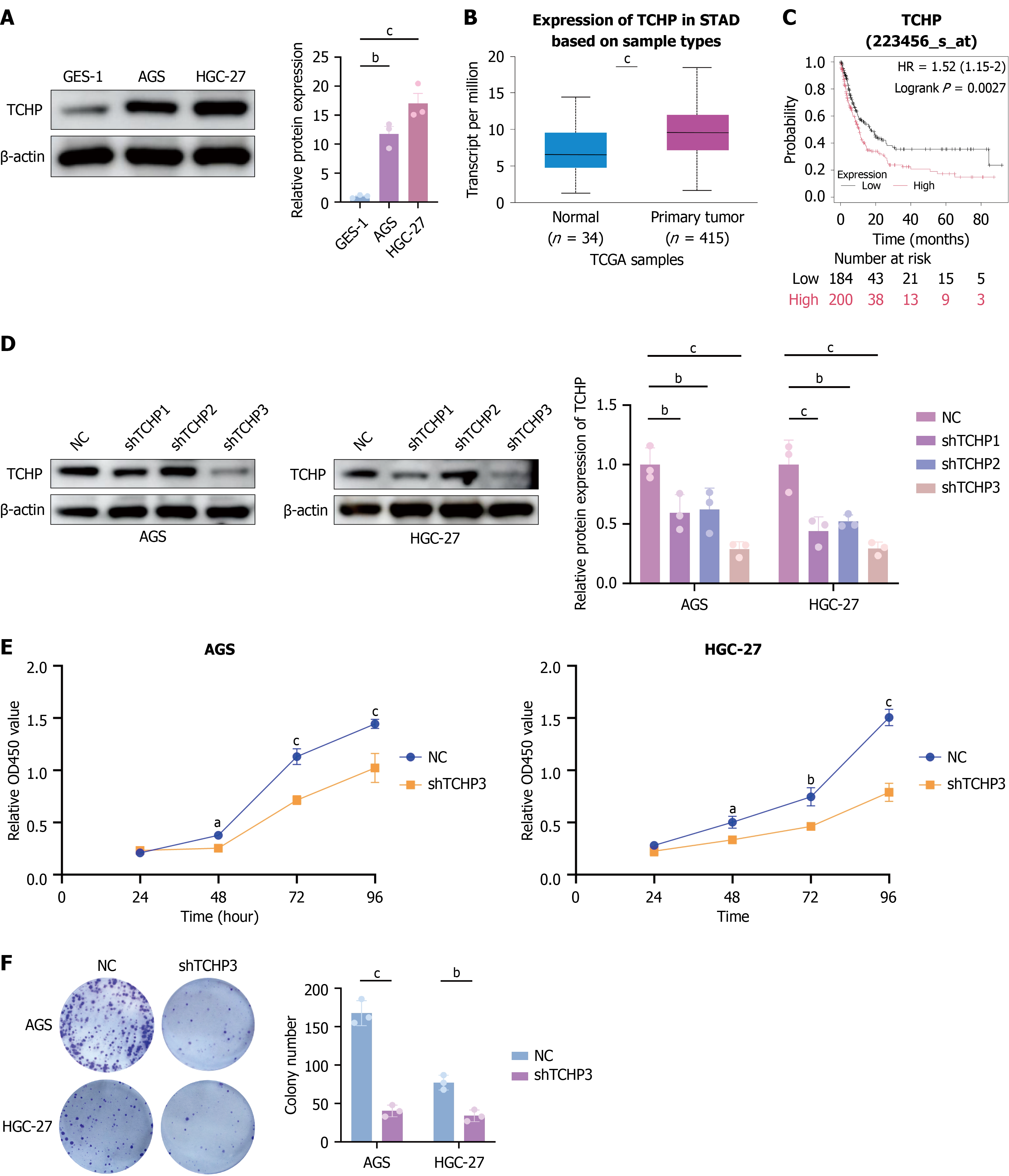
Figure 1 TCHP is upregulated in gastric cancer and promotes tumor cell proliferation.
A: Protein expression levels of TCHP in the gastric mucosal epithelial cell line (GES-1) and two gastric cancer (GC) cell lines (AGS and HGC-27) were detected by Western blotting; B: TCHP expression was significantly higher in GC tissues compared with adjacent non-tumor tissues; C: High TCHP expression was significantly associated with a lower overall survival rate in patients with GC; D: Western blotting confirmed the knockdown efficiency of TCHP in AGS and HGC-27 cells transfected with shTCHP3, compared to the negative control group; E: Cell proliferation following TCHP knockdown was assessed in AGS and HGC-27 cells using CCK-8 assay; F: Cell proliferation following TCHP knockdown was assessed in AGS and HGC-27 cells using colony formation assay. Data are presented as mean ± SD of three independent experiments. aP < 0.05; bP < 0.01; cP < 0.001. NC: Negative control; STAD: Stomach adenocarcinoma; TCGA: The Cancer Genome Atlas.
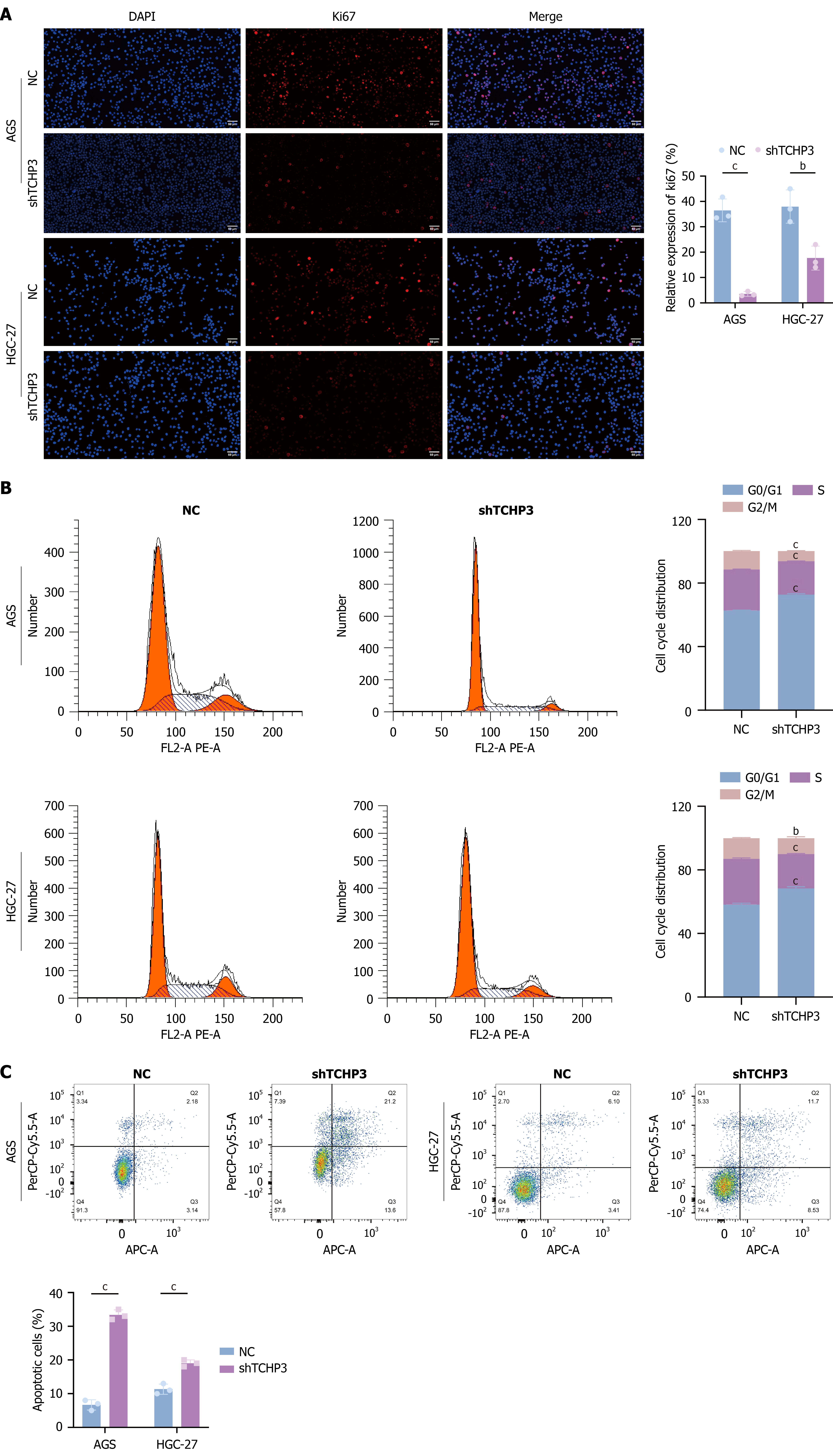
Figure 2 TCHP knockdown suppresses gastric cancer cell proliferation by inducing G1 phase cell cycle arrest and apoptosis.
A: Effect of TCHP knockdown on cell proliferation was assessed by Ki67 immunofluorescence staining; B: Analysis by flow cytometry showed that TCHP knockdown induced G1 phase cell cycle arrest in AGS and HGC-27 cells; C: Apoptosis in AGS and HGC-27 cells following TCHP knockdown was analyzed using flow cytometry. Data are presented as mean ± SD based on three independent experiments. bP < 0.01; cP < 0.001. NC: Negative control.
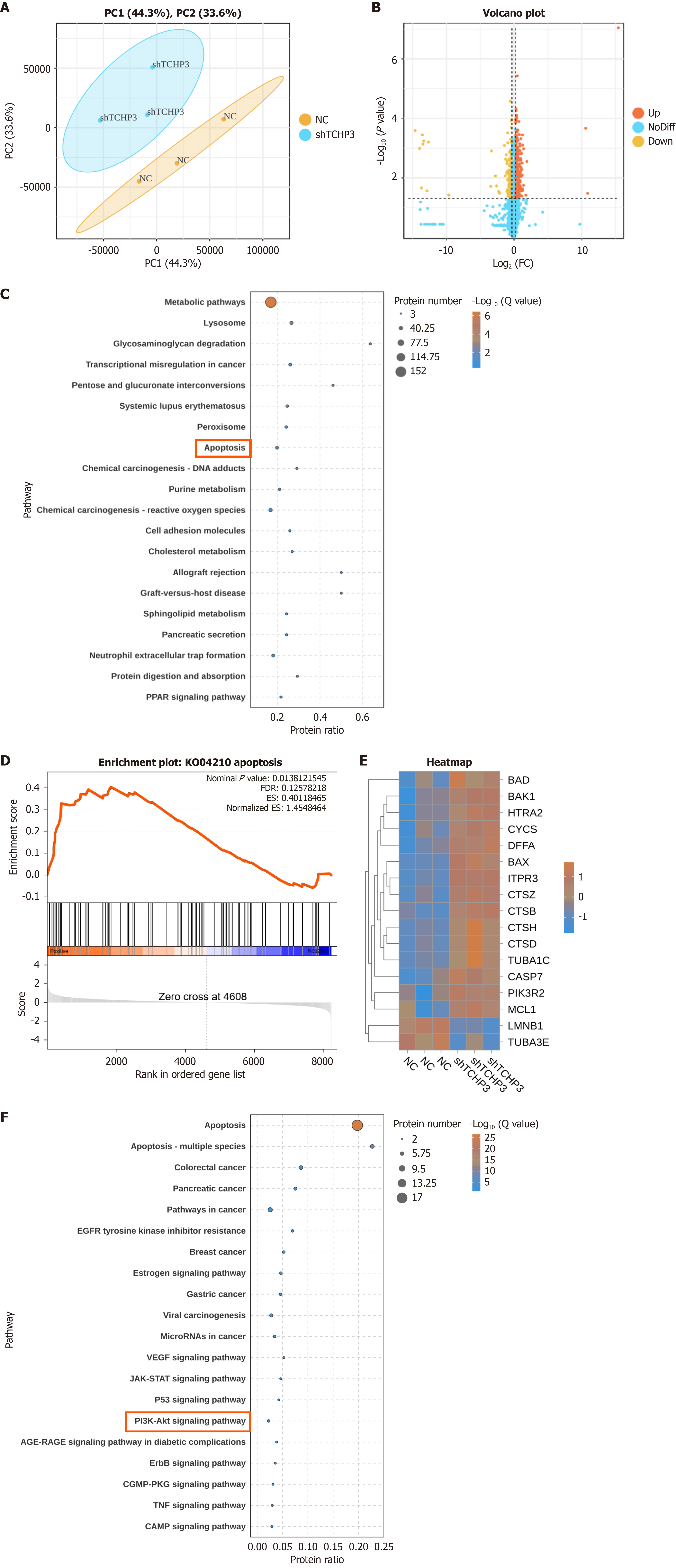
Figure 3 Proteomic analysis identifies differentially expressed proteins following TCHP knockdown.
A: Principal component analysis demonstrates distinct clustering between negative and shTCHP3 groups; B: Volcano plot shows the distribution of differentially expressed proteins (DEPs); C: Kyoto Encyclopedia of Genes and Genomes (KEGG) pathway analysis reveals significant enrichment in metabolic and apoptosis pathways; D: Gene set enrichment analysis indicates a significant upregulation of the apoptosis pathway in TCHP-knockdown gastric cancer cells; E: A heatmap shows the expression levels of key DEPs in the apoptosis pathway; F: KEGG secondary enrichment analysis identifies significant enrichment of proteins in the PI3K/AKT pathway.
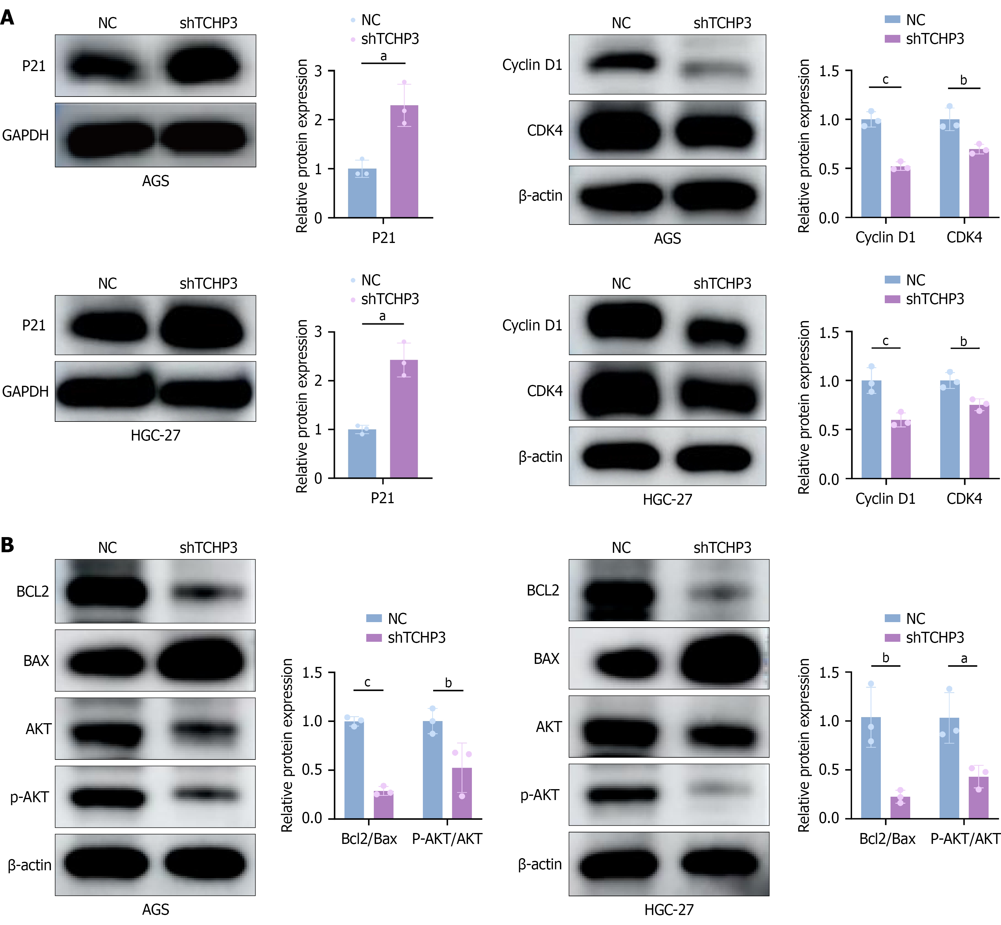
Figure 4 TCHP knockdown modulates the expression of cell cycle- and apoptosis-related proteins and affects associated signaling pathways.
A: Western blotting shows that TCHP knockdown upregulates p21 and downregulates cyclin D1 and CDK4. This pattern indicates the induction of G1 phase cell cycle arrest; B: Western blotting of AKT signaling and apoptosis-related proteins shows that TCHP knockdown reduces p-AKT and BCL2 expression, while increasing BAX levels. These changes suggest an inhibition of AKT-mediated survival signaling and promotion of apoptosis. Data are presented as mean ± SD from three independent experiments. aP < 0.05; bP < 0.01; cP < 0.001. NC: Negative control.
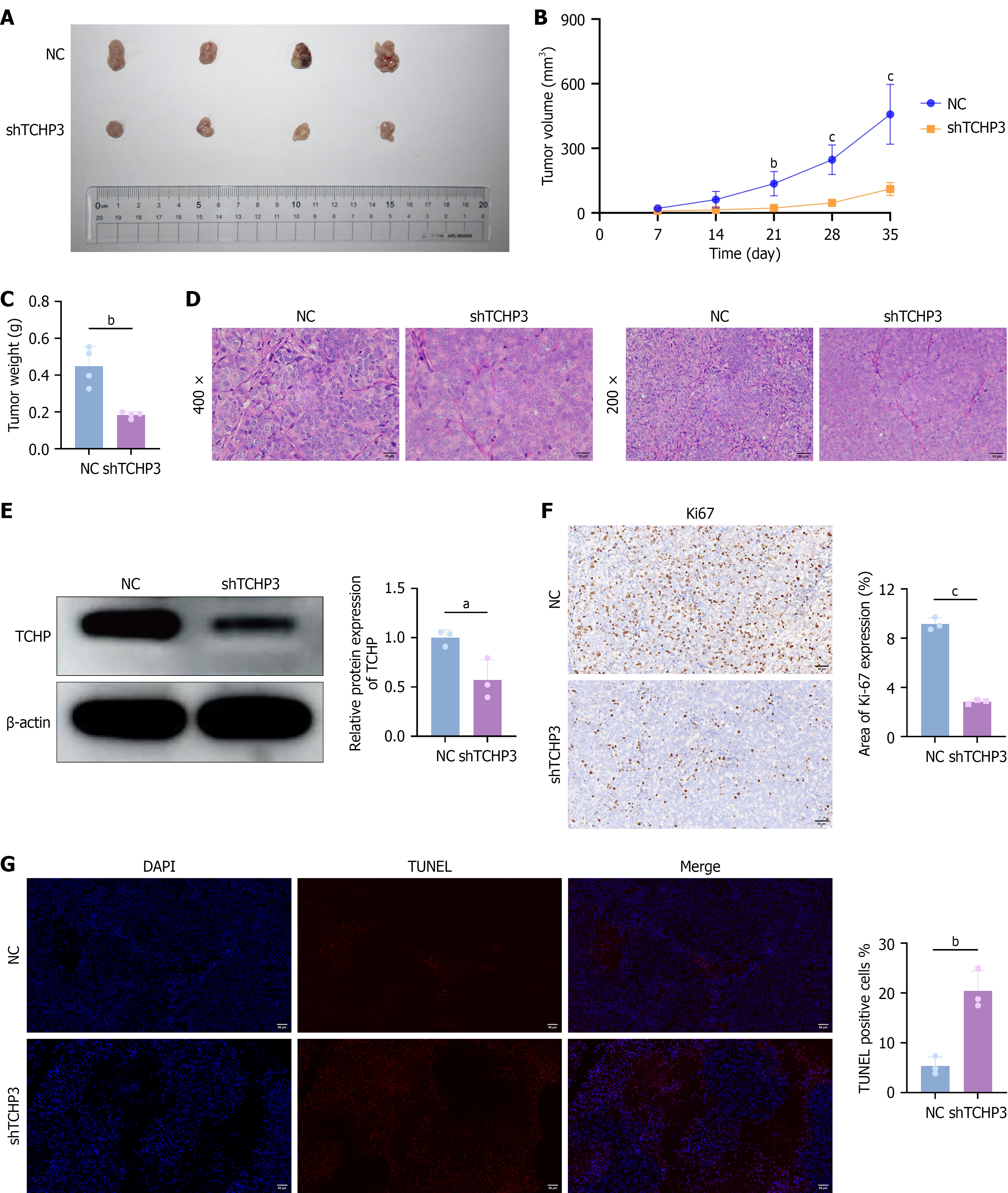
Figure 5 TCHP knockdown suppresses gastric cancer cell growth in vivo.
A: Tumor volumes in mouse xenograft models were compared between negative control (NC) and shTCHP3 groups; B: Tumor growth curves showed the tumor volumes measured every seven days; C: Final tumor weights were significantly lower in the shTCHP3 group compared to the NC group; D: Representative images of H&E staining of tumor tissues; E: Western blotting confirmed the knockdown of TCHP in the harvested tumors; F: The expression of Ki67 protein was detected by immunohistochemistry; G: Apoptosis in the subcutaneous xenograft model was assessed by the TUNEL assay. Data are presented as mean ± SD; n = 4 mice per group. aP < 0.05; bP < 0.01; cP < 0.001. NC: Negative control.
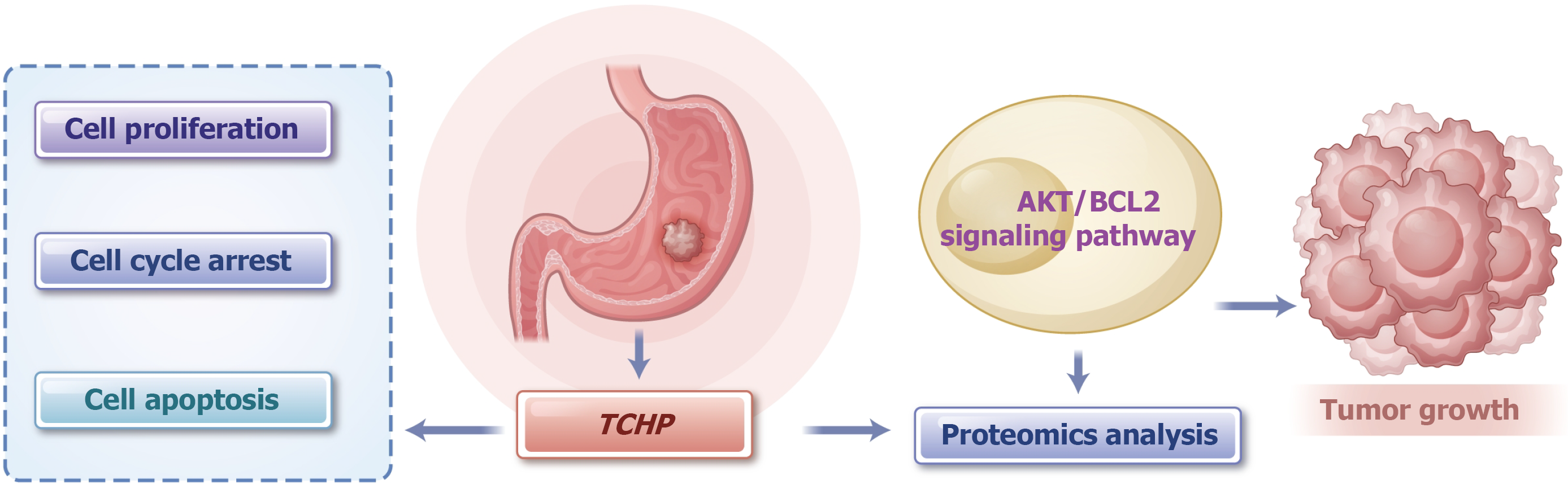
Figure 6
Schematic diagram of the mechanisms by which TCHP regulates the proliferation and apoptosis of gastric cancer cells through the AKT/BCL2 signaling pathway.
- Citation: Wang Y, Zhang TY, Shen YF, Shen N, Zhang ZW. TCHP regulates the proliferation and apoptosis of gastric cancer cells via AKT/BCL2 signaling pathway. World J Gastrointest Oncol 2026; 18(8): 116937
- URL: https://www.wjgnet.com/1948-5204/full/v18/i8/116937.htm
- DOI: https://dx.doi.org/10.4251/wjgo.116937