©The Author(s) 2025.
World J Gastrointest Oncol. Aug 15, 2025; 17(8): 105818
Published online Aug 15, 2025. doi: 10.4251/wjgo.v17.i8.105818
Published online Aug 15, 2025. doi: 10.4251/wjgo.v17.i8.105818
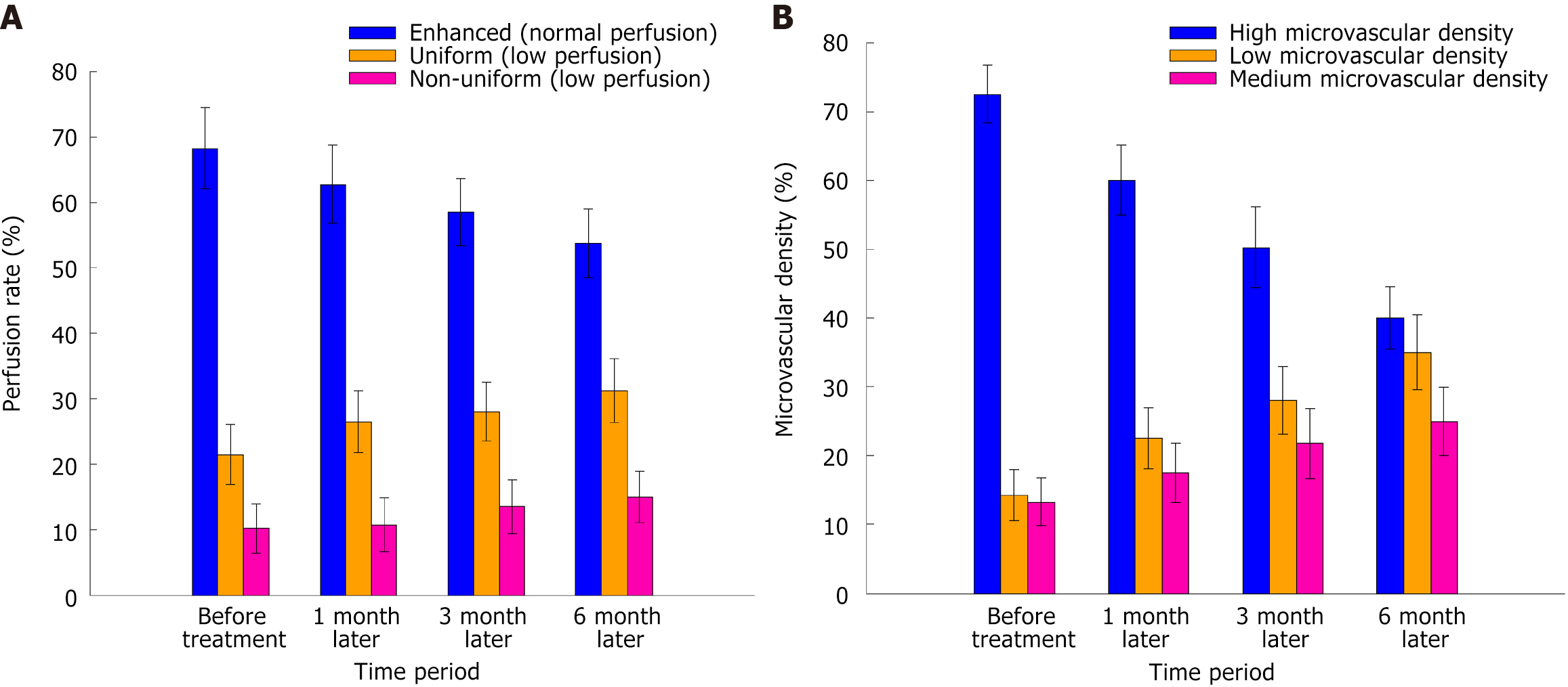
Figure 1 Different performance.
A Differences in tumor blood perfusion patterns at various stages of treatment; B: Differences in tumor microvessel density in patients across different periods.
Figure 2 Ultrasound image illustrating a typical case.
A: It depicts a standard instance of preablation ultrasonography using a low-frequency convex array probe. The arterial phase of the solid mass at the proximal peritoneum of hepatic S4 exhibited rapid enhancement within a brief duration, implying robust neoangiogenesis and ample blood supply, characteristic of tumor malignancy; B: High-frequency line array probe; C: Low-frequency convex array probe. They illustrate a typical postablation ultrasonography scenario. In the arterial phase, the solid mass at the proximal peritoneum of hepatic S4 displayed no enhancement, signifying a near-complete cessation of blood flow in the tumor region post-treatment without evident perfusion, indicating a favorable therapeutic outcome.
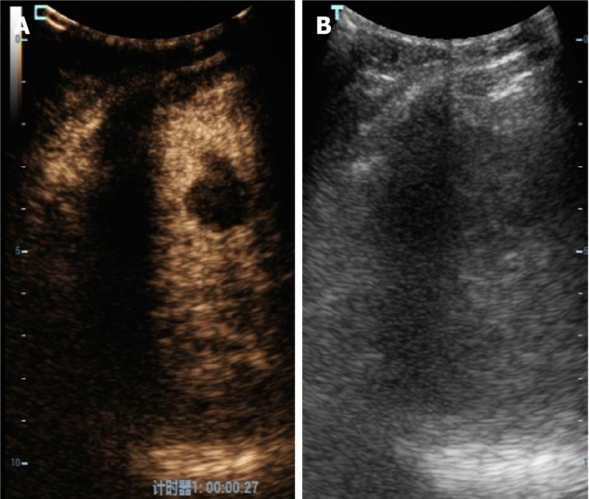
Figure 3 Typical cases of perfusion before and after treatment.
A: It depicts the preoperative contrast-enhanced ultrasound of the ablation, revealing pronounced enhancement in the arterial phase of the solid liver S8 mass, suggesting sufficient blood supply to the tumor and significantly enhanced perfusion, typically observed in malignant tumors; B: The arterial phase of the solid liver S8 mass exhibits no enhancement on contrast-enhanced ultrasound post-ablation, indicating complete cessation of blood flow in the tumor region post-treatment, absence of evident perfusion, and either remission or cure of the tumor.
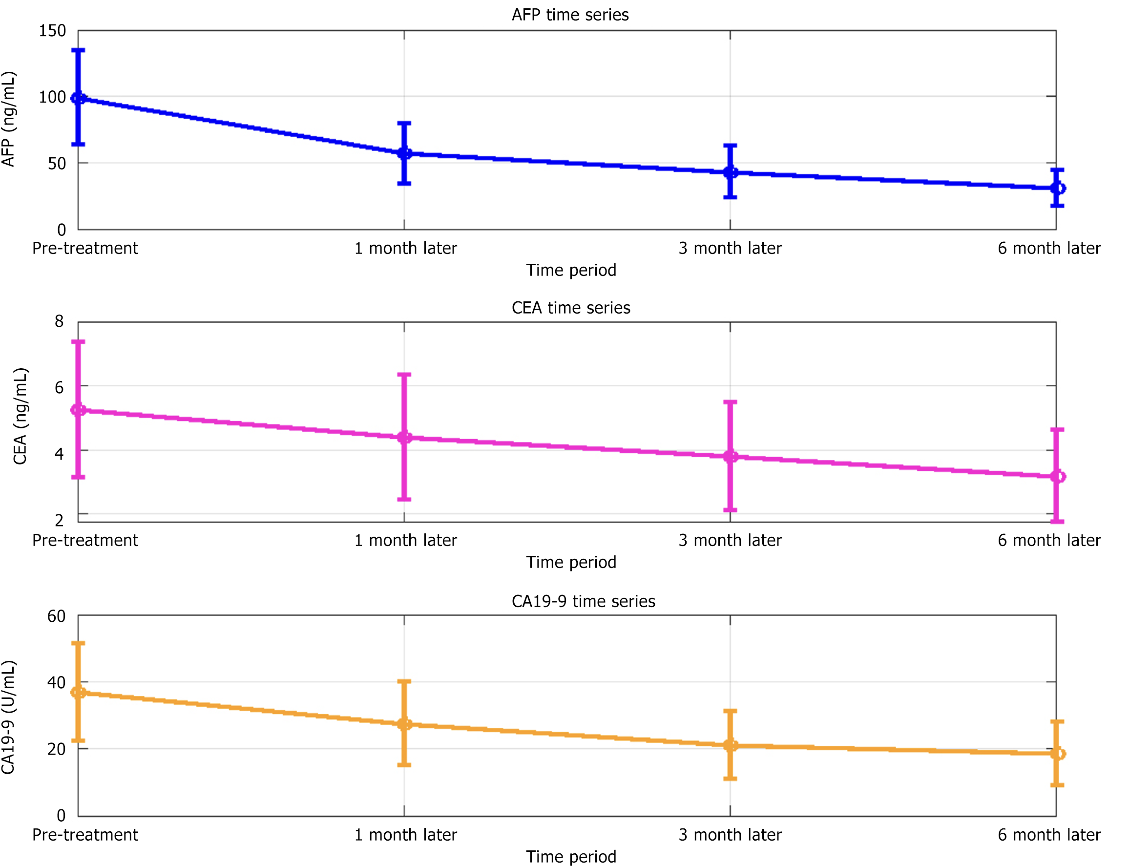
Figure 4 Changes of tumor markers in patients at different periods.
AFP: Alpha-fetoprotein; CA 19-9: Carbohydrate antigen 19-9; CEA: Carcinoembryonic antigen.
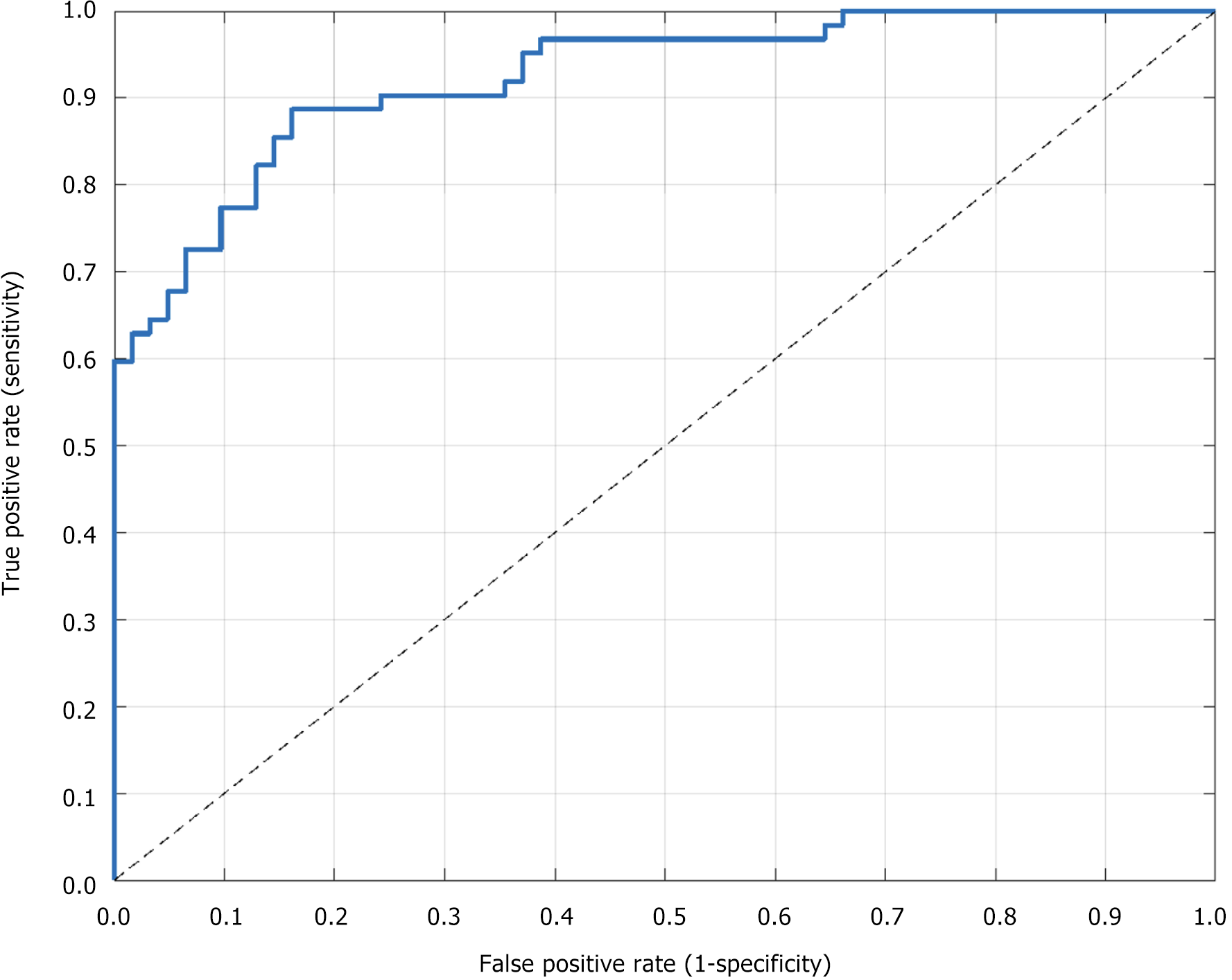
Figure 5 Receiver operating characteristic curve depicting the diagnostic accuracy of contrast-enhanced ultrasonography.
- Citation: Chen LP, Dong Y, He JG, Yang QQ, Hu ZW. Contrast-enhanced ultrasound in evaluating the curative effect of interventional therapy in patients with liver cancer. World J Gastrointest Oncol 2025; 17(8): 105818
- URL: https://www.wjgnet.com/1948-5204/full/v17/i8/105818.htm
- DOI: https://dx.doi.org/10.4251/wjgo.v17.i8.105818


