©The Author(s) 2025.
World J Gastrointest Oncol. Jul 15, 2025; 17(7): 108650
Published online Jul 15, 2025. doi: 10.4251/wjgo.v17.i7.108650
Published online Jul 15, 2025. doi: 10.4251/wjgo.v17.i7.108650
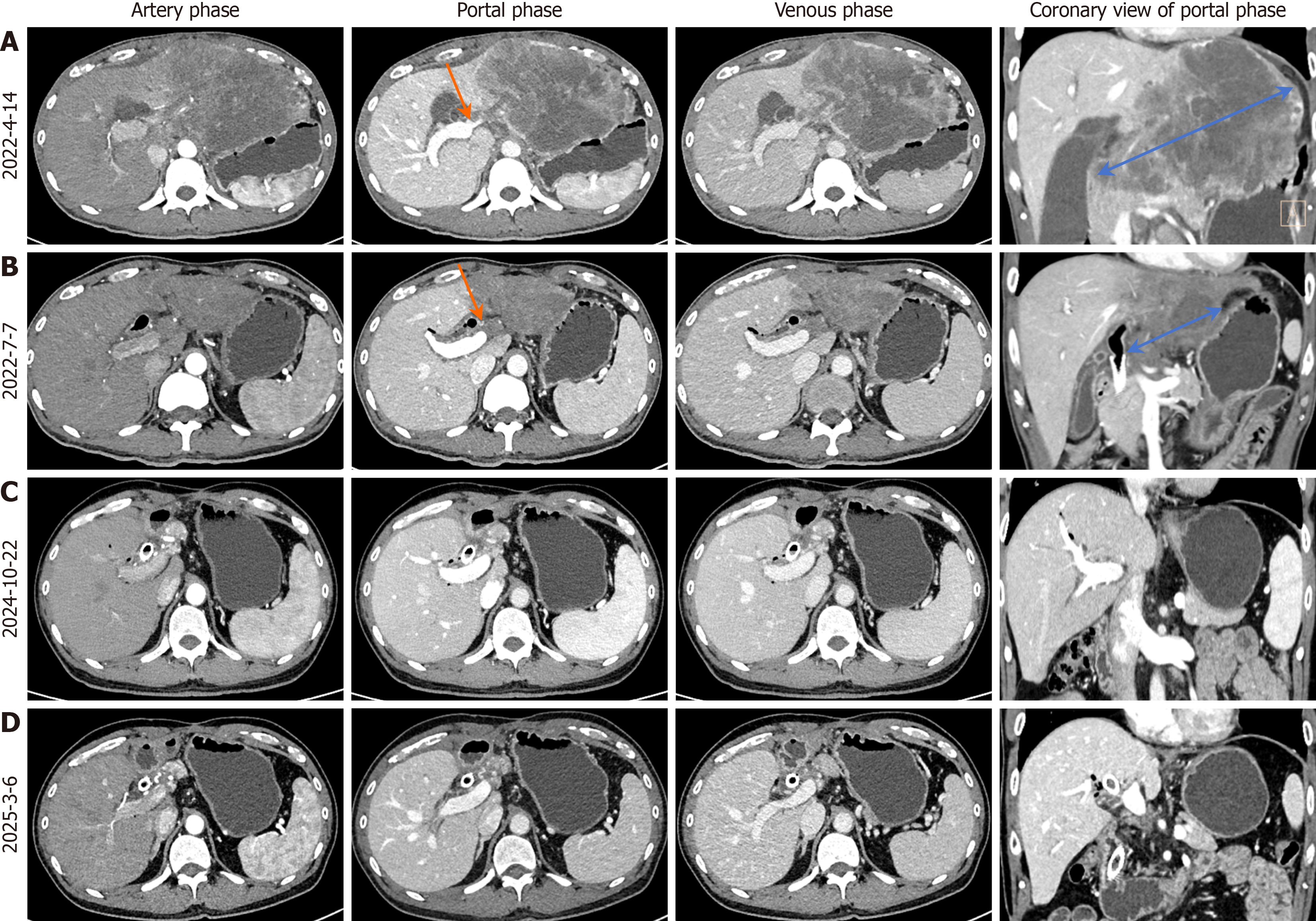
Figure 1 Serial contrast-enhanced computed tomography imaging during the treatment course.
A: Pre-chemotherapy baseline computed tomography (CT) showing a 13-cm hypodense mass (blue arrow) with heterogeneous enhancement in the left hepatic lobe, exhibiting mild to moderate peripheral enhancement and left portal branch encasement (orange arrow); B: Follow-up CT after 3 cycles of combined therapy demonstrating a significant reduction in tumor diameter to 6.5 cm (blue arrow), with attenuated enhancement compared to baseline and alleviated left portal branch involvement (orange arrow); C: Post-resection CT at 24-month follow-up revealing no evidence of tumor recurrence; D: Post-resection CT at 29-month follow-up confirming the absence of tumor recurrence.
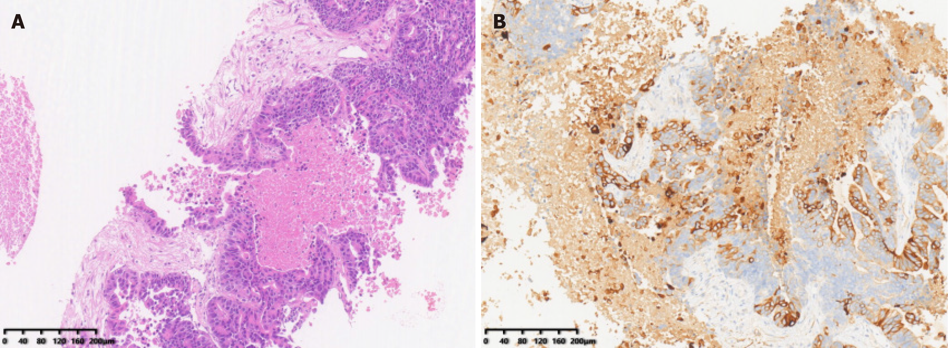
Figure 2 Histopathological and immunohistochemical features of the liver tumor biopsy.
A: Hematoxylin and eosin staining (× 10) reveals adenocarcinoma with cuboidal tumor cells arranged in glandular patterns; B: Immunohistochemical staining for CK7 is positive, confirming biliary differentiation.
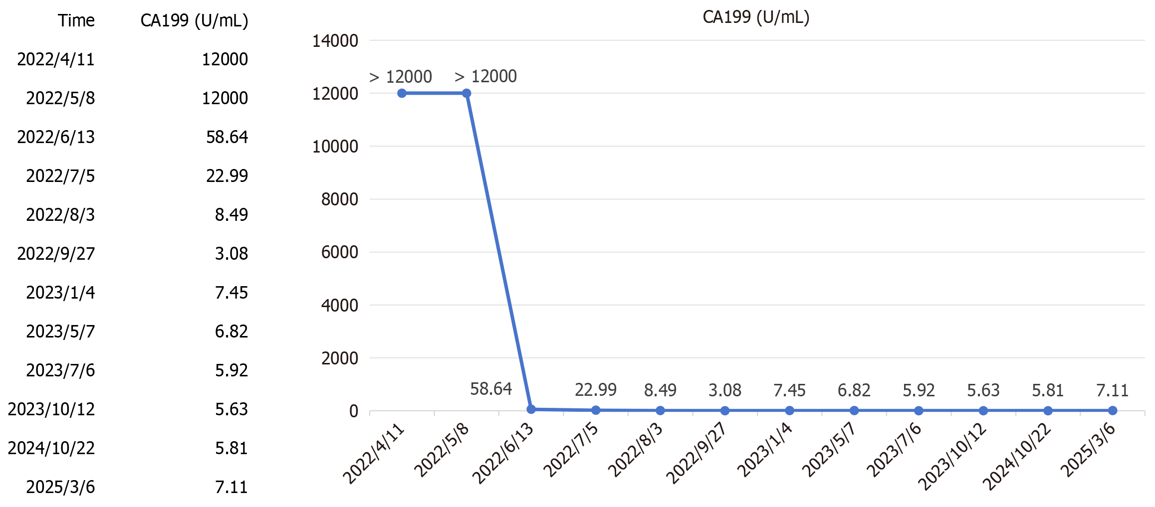
Figure 3
Serial carbohydrate antigen 19-9 monitoring during treatment shows that carbohydrate antigen 19-9 decreased significantly after two cycles of combined therapy, returned to the normal range after three cycles, and remained within the normal range during postoperative follow-up.
Figure 4
Contrast-enhanced computed tomography imaging of the hepatic abscess, demonstrating diffuse hepatic parenchymal edema, with a characteristic targetoid peripheral rim enhancement encircling the hypodense abscess wall and a central non-enhancing hypodense area indicative of liquefactive necrosis.
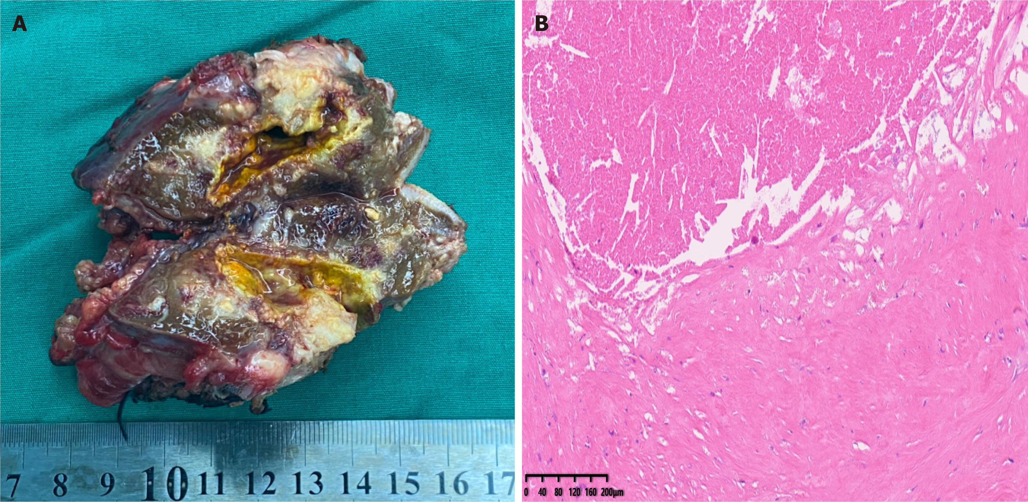
Figure 5 Tumor pathology.
A: Gross resected tumor specimen; B: Hematoxylin and eosin staining, × 10. Tissue was necrotic; no residual viable tumor was detected.
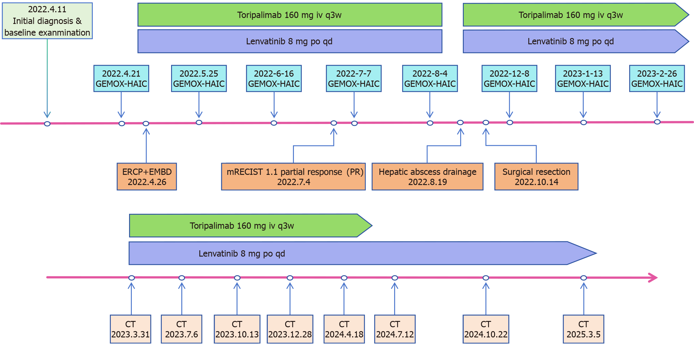
Figure 6 Clinical timeline depicting the sequence of initial diagnosis, implementation of GEMOX-HAIC combined with lenvatinib and toripalimab therapy, complication management, subsequent surgical resection, and post-operative follow-up.
CT: Computed tomography.
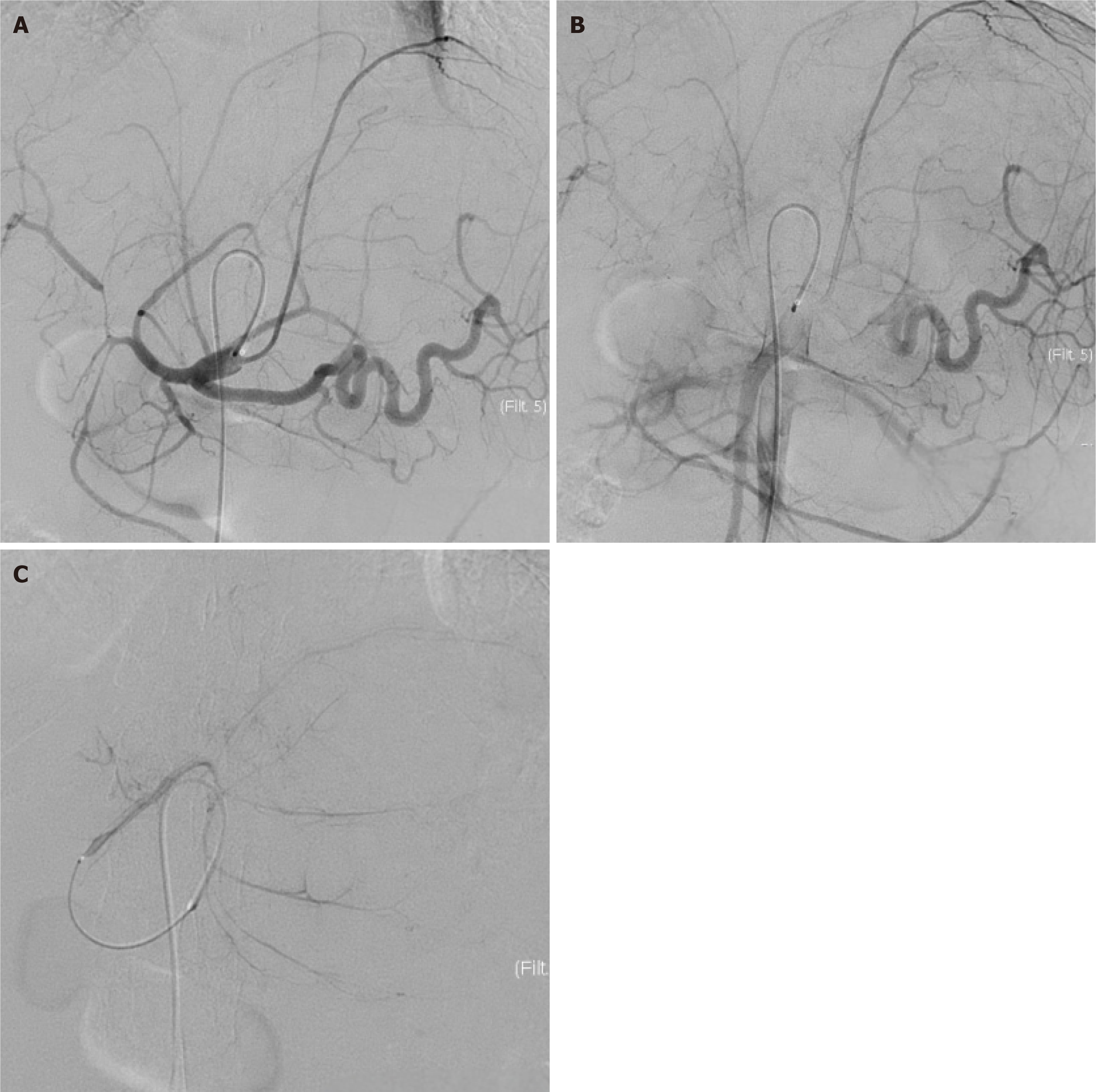
Figure 7 Pre-chemotherapy hepatic arteriography.
A and B: The left hepatic artery is the dominant feeding artery of the tumor, demonstrating marked luminal dilation, serpentine tortuosity, and proliferative branching encircling the tumor in the early and middle arterial phases; C: Prominent tumor staining is observed in the late arterial phase during microcatheter angiography.
- Citation: Xie JP, Tang YJ, Fan YW, Huang YZ, Deng M, Zhang TZ, Li Y, Deng G, Tang D. Pathological complete response in advanced intrahepatic cholangiocarcinoma was achieved through tri-modal therapy: A case report and review of literature. World J Gastrointest Oncol 2025; 17(7): 108650
- URL: https://www.wjgnet.com/1948-5204/full/v17/i7/108650.htm
- DOI: https://dx.doi.org/10.4251/wjgo.v17.i7.108650


