©The Author(s) 2025.
World J Gastrointest Oncol. Jul 15, 2025; 17(7): 107971
Published online Jul 15, 2025. doi: 10.4251/wjgo.v17.i7.107971
Published online Jul 15, 2025. doi: 10.4251/wjgo.v17.i7.107971
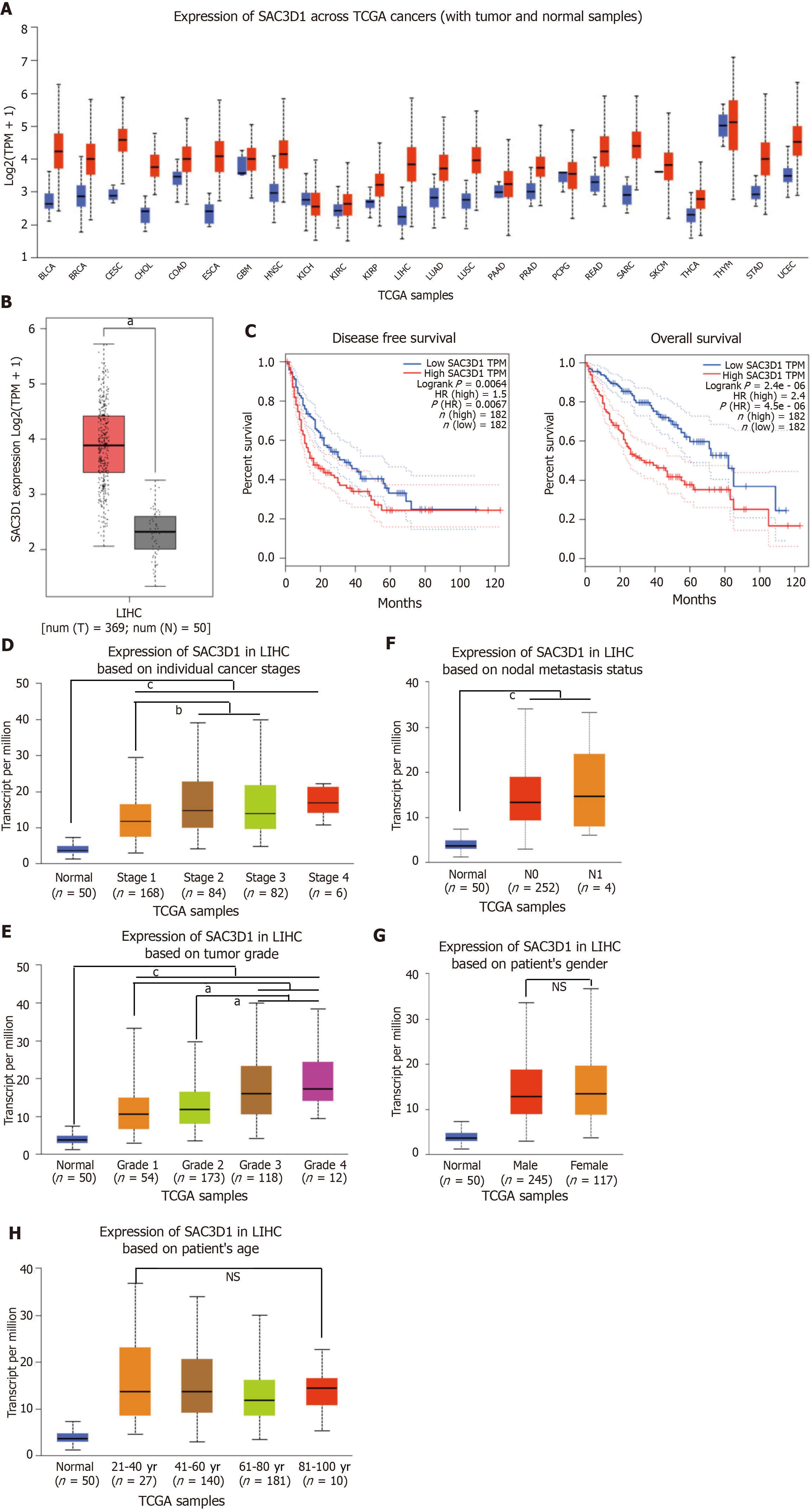
Figure 1 SAC3 domain containing 1 is upregulated in liver hepatocellular carcinoma and is associated with tumor grade and disease prognosis.
aP < 0.05, bP < 0.01, cP < 0.001. A: Analysis of SAC3 domain containing 1 (SAC3D1) expression across different cancers using UALCAN database; B: Analysis of SAC3D1 expression in liver hepatocellular carcinoma and adjacent tissues using gene expression profiling interactive analysis; C: Correlation between high and low SAC3D1 expression and overall survival and progression-free survival in liver hepatocellular carcinoma patients, as analyzed by gene expression profiling interactive analysis; D: Correlation between SAC3D1 transcription levels and tumor stages; E: Correlation between SAC3D1 transcription levels and tumor grades; F: Correlation between SAC3D1 transcription levels and lymph node metastasis; G: Correlation between SAC3D1 transcription levels and gender; H: Correlation between SAC3D1 transcription levels and age. SAC3D1: SAC3 domain containing 1; LIHC: Liver hepatocellular carcinoma; TCGA: The Cancer Genome Atlas; NS: No significance.
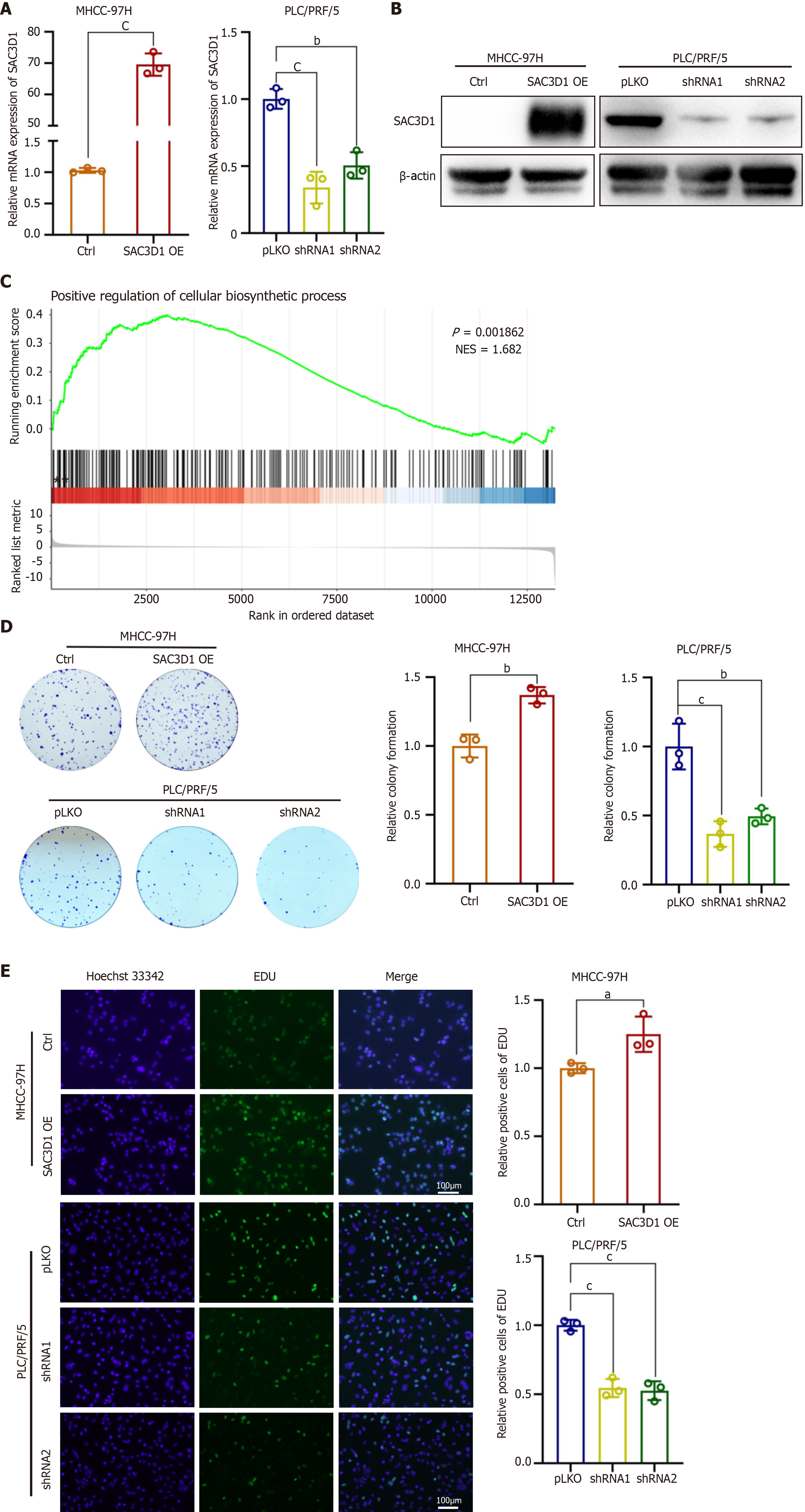
Figure 2 In vitro, SAC3 domain containing 1 promotes the proliferation of hepatocellular carcinoma cells.
aP < 0.05, bP < 0.01, cP < 0.001. A: Real-time quantitative reverse transcription polymerase chain reaction was used to detect the transcriptional levels of SAC3 domain containing 1 (SAC3D1) in cell sublines with overexpressed and knocked down SAC3D1; B: Western blotting was employed to assess the protein levels of SAC3D1 in cell sublines with overexpressed and knocked down SAC3D1; C: Gene set enrichment analysis was conducted to explore the signaling pathways affected by the upregulation of SAC3D1 expression; D: Colony formation assays were conducted to validate the impact of SAC3D1 on the proliferative capacity of hepatocellular carcinoma cells; E: The 5-ethynyl-2′-deoxyuridine assay was performed to evaluate the impact of SAC3D1 overexpression on hepatocellular carcinoma cell proliferation, bar = 100 μm. SAC3D1: SAC3 domain containing 1; EdU: 5-ethynyl-2’-deoxyuridine.
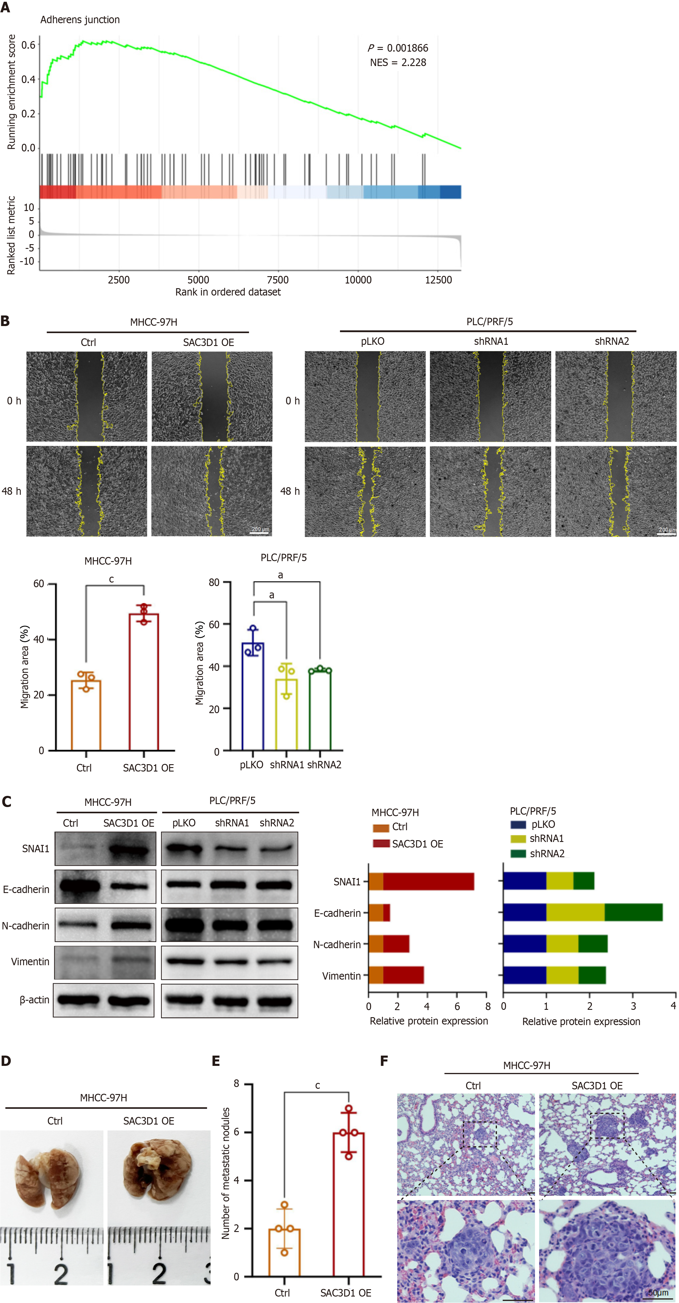
Figure 3 SAC3 domain containing 1 promotes metastasis of hepatocellular carcinoma cells in vitro and in vivo.
aP < 0.05, cP < 0.001. A: Gene set enrichment analysis was conducted to explore the signaling pathways affected by the upregulation of SAC3 domain containing 1 (SAC3D1) expression; B: The wound healing assay was utilized to validate the effect of SAC3D1 overexpression on hepatocellular carcinoma cell migration, bar = 200 μm; C: Western blotting results demonstrate that the expression level of SAC3D1 modulates the expression of migration-related proteins in hepatocellular carcinoma cells; D: Metastatic tumor nodules in lungs of two groups of nude mice; E: The numbers of metastatic tumor nodules per field of view in lungs of two groups; F: The metastatic tumor nodules in lungs of nude mice by hematoxylin and eosin staining, bar = 50 μm. SAC3D1: SAC3 domain containing 1; SNAI1: Snail family transcriptional repressor 1.
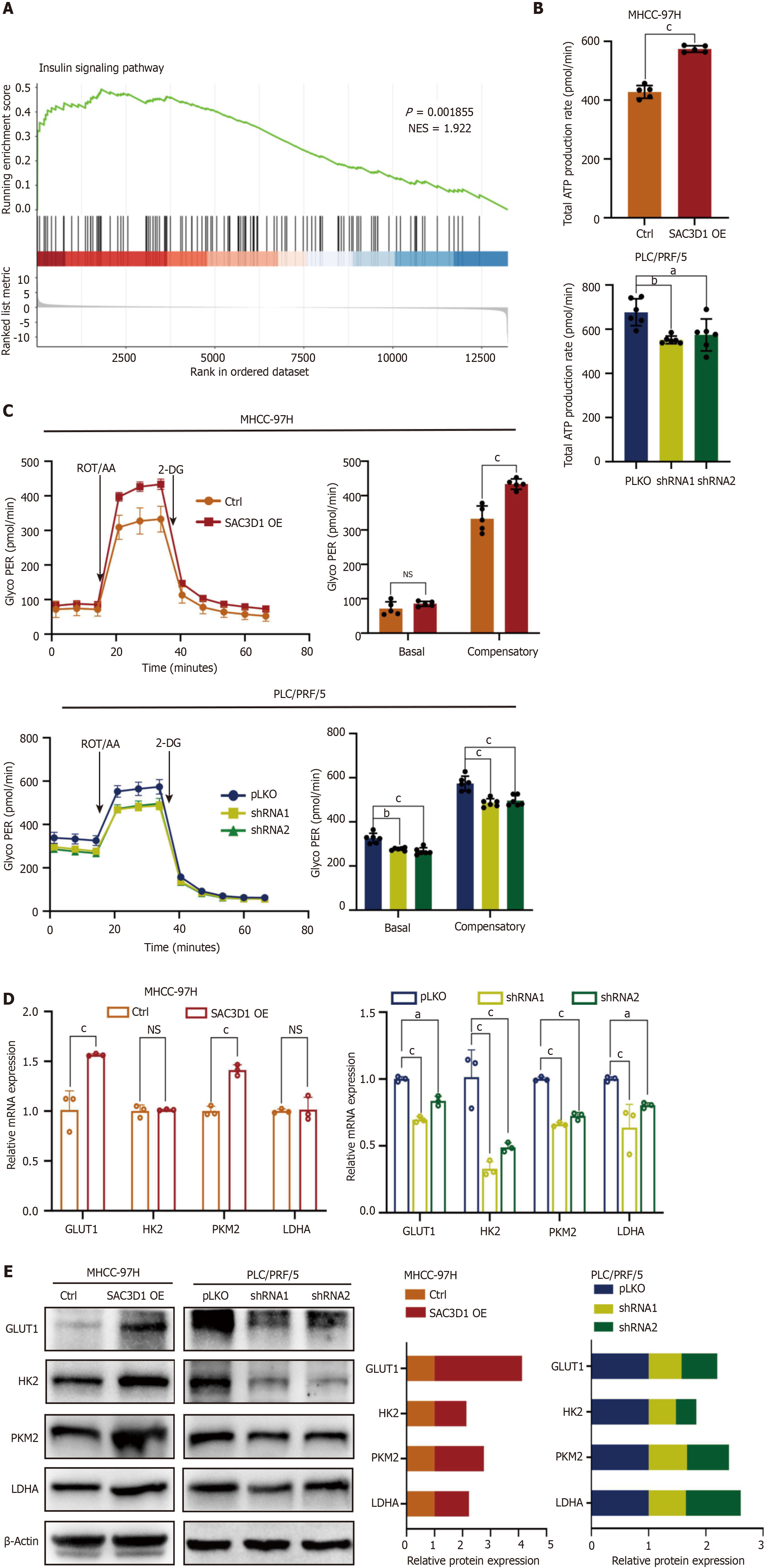
Figure 4 SAC3 domain containing 1 enhances the glycolysis and total adenosine triphosphate production in hepatocellular carcinoma cells.
aP < 0.05, bP < 0.01, cP < 0.001. A: Gene set enrichment analysis indicated that SAC3 domain containing 1 (SAC3D1) promoted activation of the insulin signaling pathway; B: The Seahorse XFe96 assay was used to assess the impact of SAC3D1 on the total adenosine triphosphate production rate in hepatocellular carcinoma cells; C: The Seahorse XFe96 assay was utilized to evaluate the effect of SAC3D1 on the glycolytic rate in hepatocellular carcinoma cells; D: Real-time quantitative reverse transcription polymerase chain reaction was performed to examine the influence of SAC3D1 on the transcription levels of glycolysis-related genes; E: Western blotting was conducted to assess the effect of SAC3D1 on the expression of glycolysis-related proteins. SAC3D1: SAC3 domain containing 1; ROT/AA: Rotenone and antimycin A; NS: No significance; 2-DG: 2-deoxyglucose; GLUT1: Glucose transporter 1; HK2: Hexokinase 2; PKM2: Pyruvate kinase M2; LDHA: Lactate dehydrogenase A.
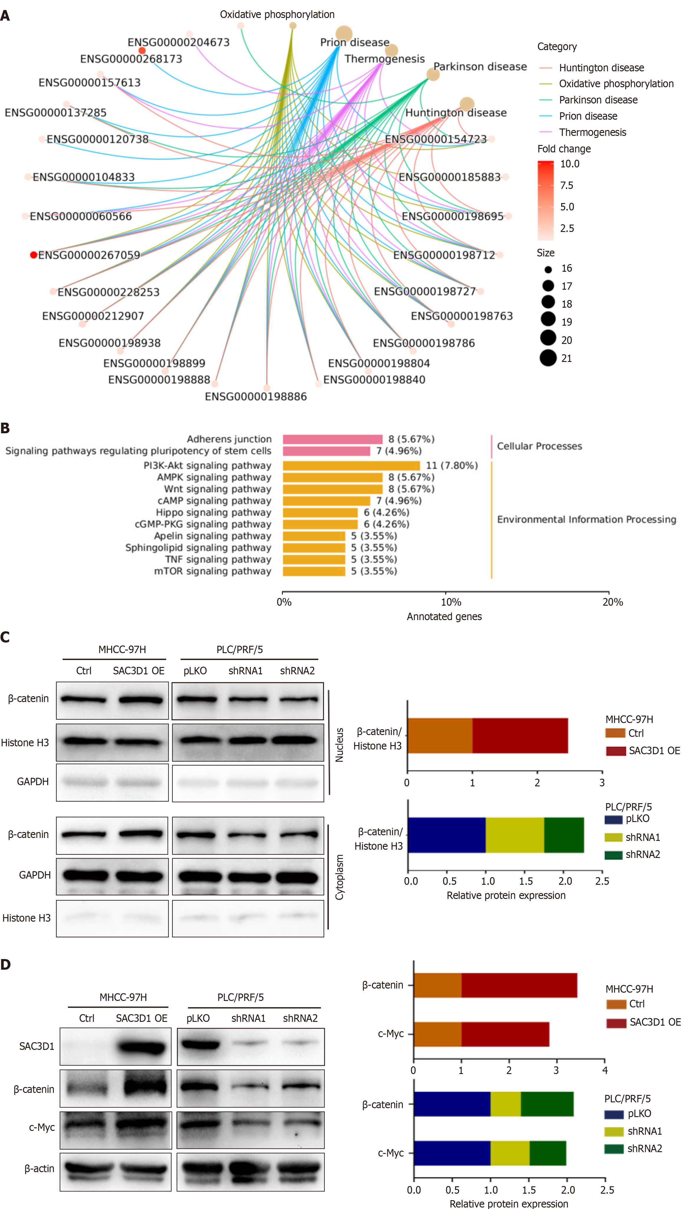
Figure 5 SAC3 domain containing 1 participates in the energy metabolism reprogramming of hepatocellular carcinoma cells through the β-Catenin/c-Myc signaling axis.
A: Kyoto Encyclopedia of Genes and Genomes enrichment network diagram of differential genes; B: Kyoto Encyclopedia of Genes and Genomes clustering analysis diagram; C: Nuclear-cytoplasmic fractionation and western blotting were performed to assess β-Catenin protein levels in the nuclear and cytoplasmic compartments; D: Western blotting analysis of the correlation between SAC3 domain containing 1 and the expression of β-Catenin and c-Myc. SAC3D1: SAC3 domain containing 1.
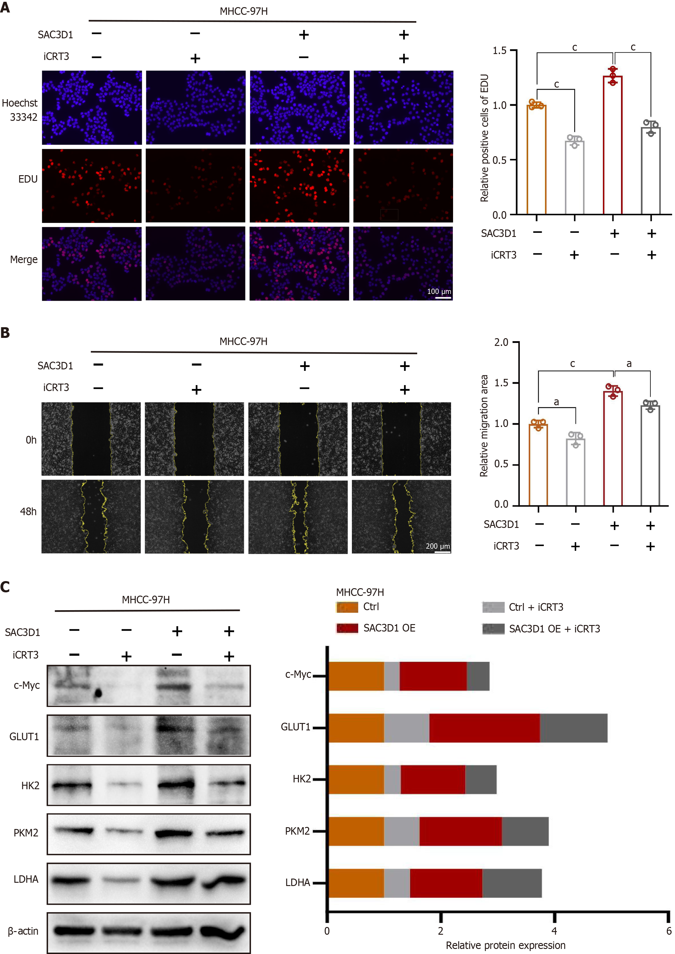
Figure 6 The inhibitor of β-Catenin, iCRT3, can attenuate the cellular phenotypes induced by SAC3 domain containing 1 overexpression.
aP < 0.05, cP < 0.001. A: The iCRT3 antagonizes the enhanced cell proliferation ability caused by SAC3 domain containing 1 overexpression, bar = 100 μm; B: The iCRT3 antagonizes the enhanced cell migration ability caused by SAC3 domain containing 1 overexpression, bar = 200 μm; C: Western blotting analysis of the effects of the β-Catenin antagonist iCRT3 on the expression of c-Myc, glucose transporter 1, hexokinase 2, pyruvate kinase M2 and lactate dehydrogenase A. SAC3D1: SAC3 domain containing 1; EdU: 5-ethynyl-2’-deoxyuridine; GLUT1: Glucose transporter 1; HK2: Hexokinase 2; PKM2: Pyruvate kinase M2; LDHA: Lactate dehydrogenase A.
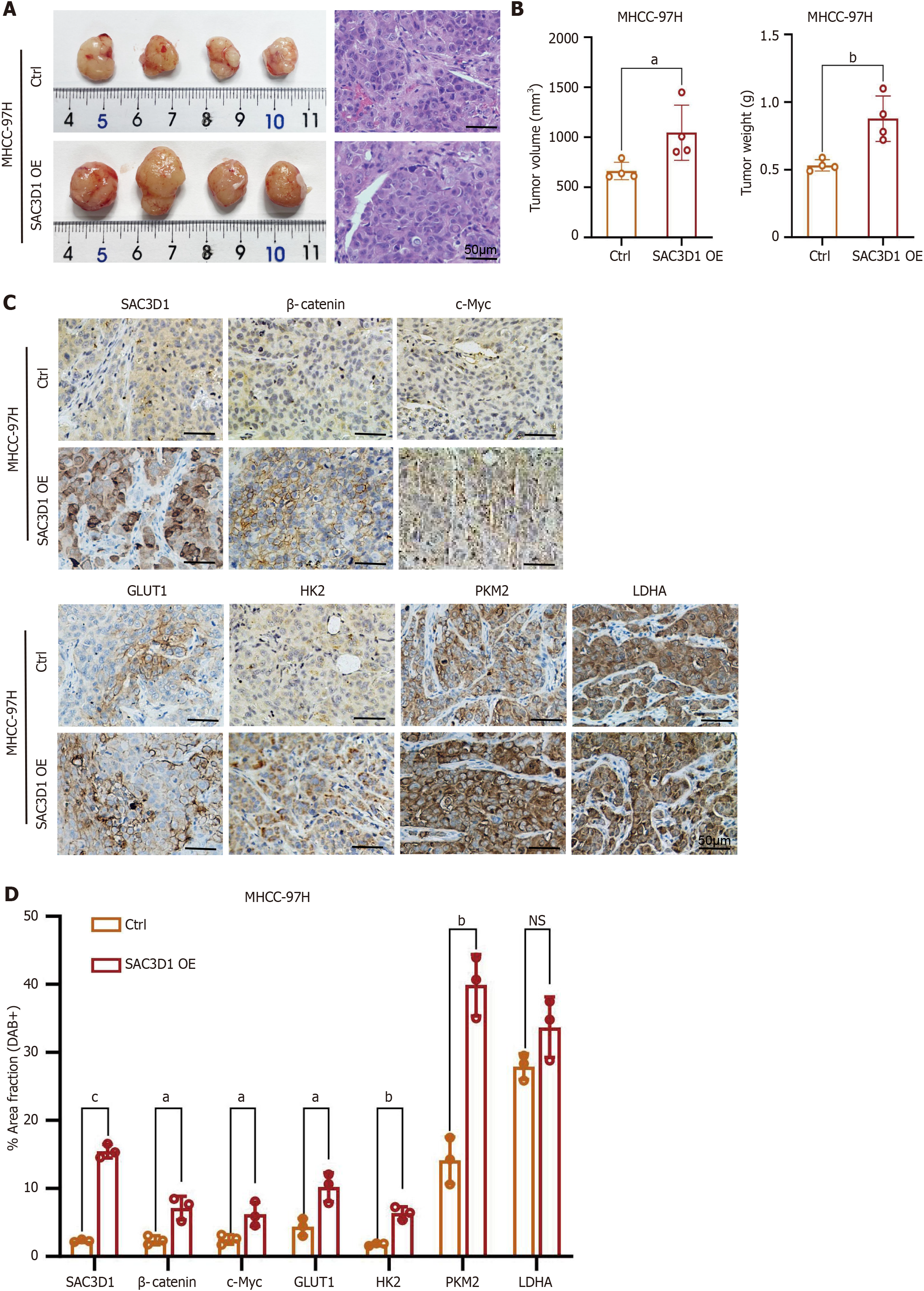
Figure 7 In vivo, SAC3 domain containing 1 significantly promotes the progression of hepatocellular carcinoma.
aP < 0.05, bP < 0.01, cP < 0.001. A: Xenograft tumor of MHCC-97H SAC3 domain containing 1 overexpression and MHCC-97H control (Ctrl) cells and hematoxylin and eosin staining, bar = 50 μm; B: Statistical analysis of xenograft tumor volume and tumor weight; C and D: Immunohistochemistry detection and statistical analysis of the expression and positive rates of SAC3 domain containing 1, β-Catenin, c-Myc, glucose transporter 1, hexokinase 2, pyruvate kinase M2 and lactate dehydrogenase A, bar = 50 μm. OE: Over expression; NS: No significance; SAC3D1: SAC3 domain containing 1;GLUT1: Glucose transporter 1; HK2: Hexokinase 2; PKM2: Pyruvate kinase M2; LDHA: Lactate dehydrogenase A.
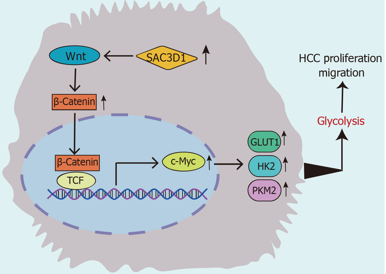
Figure 8 Molecular mechanism by which SAC3 domain containing 1 promotes the progression of hepatocellular carcinoma.
In hepatocellular carcinoma cells, the upregulated expression of SAC3 domain containing 1 enhances the expression of glycolysis-related genes by regulating the β-Catenin/c-Myc signaling axis, thereby increasing the cellular glycolytic capacity and adenosine triphosphate production rate to meet the demands of large-scale biosynthesis and proliferation in cancer cells, ultimately promoting the progression of hepatocellular carcinoma. SAC3D1: SAC3 domain containing 1; TCF: T-cell factor; GLUT1: Glucose transporter 1; HK2: Hexokinase 2; PKM2: Pyruvate kinase M2; LDHA: Lactate dehydrogenase A.
- Citation: Lin XJ, Tang EJ, Sun B, Wang AL, Chen Y, Chen L, Xue YY, Li AJ, Liu CY. SAC3 domain containing 1 intervention in energy metabolism reprogramming assists in the progression of hepatocellular carcinoma. World J Gastrointest Oncol 2025; 17(7): 107971
- URL: https://www.wjgnet.com/1948-5204/full/v17/i7/107971.htm
- DOI: https://dx.doi.org/10.4251/wjgo.v17.i7.107971


