©The Author(s) 2025.
World J Gastrointest Oncol. Jul 15, 2025; 17(7): 106701
Published online Jul 15, 2025. doi: 10.4251/wjgo.v17.i7.106701
Published online Jul 15, 2025. doi: 10.4251/wjgo.v17.i7.106701
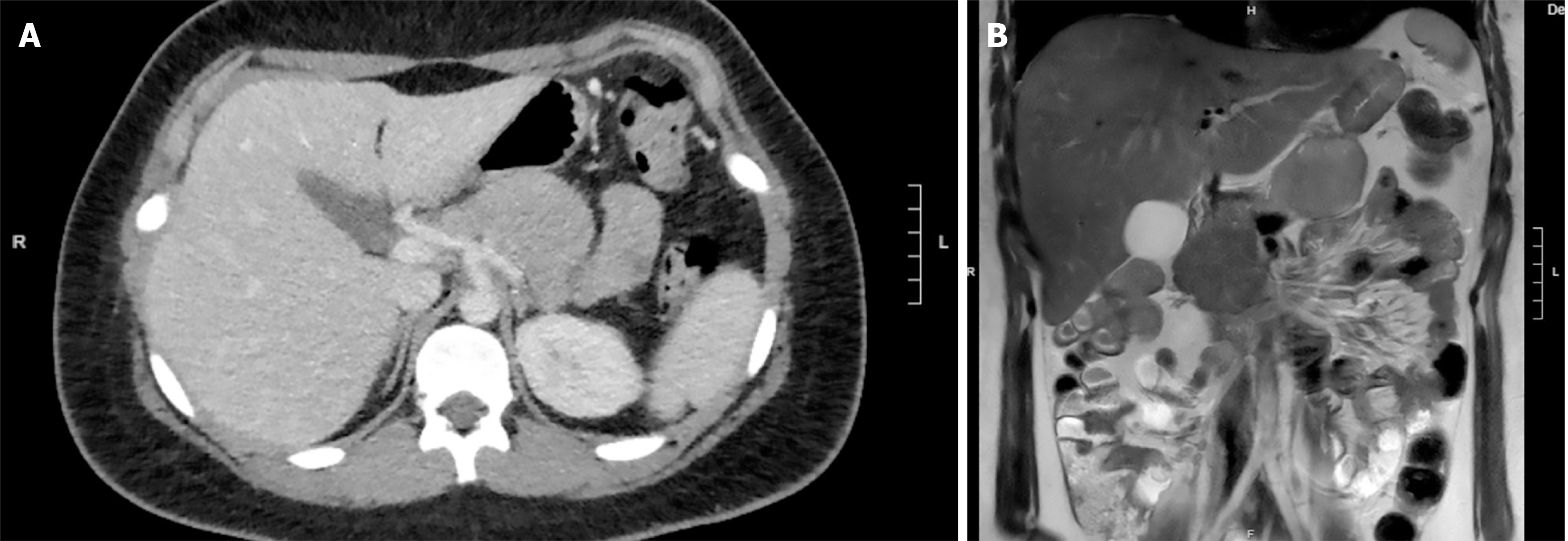
Figure 1 Computed tomography axial and magnetic resonance imaging coronal view demonstrating pre-surgical pancreatic mass.
A: Computed tomography axial view demonstrating pre-surgical pancreatic mass; B: Magnetic resonance imaging coronal view demonstrating pre-surgical pancreatic mass.
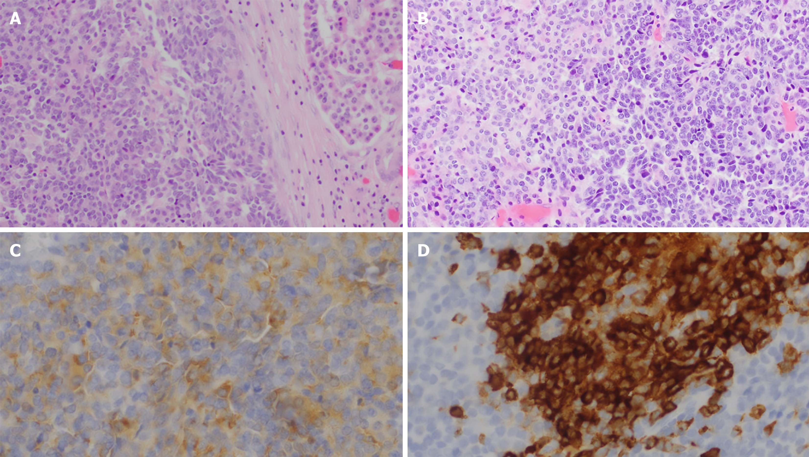
Figure 2 Histological slides from her tumor can be appreciated.
A: Hematoxylin and eosin stain demonstrating the interface between tumor (right) and a pancreatic islet with adjacent duct (left). Tumor has suggestion of rosette formation; B: Hematoxylin and eosin stain demonstrating the immature small round blue cell component (lower right) and acinar component on (upper left); C: Immunohistochemistry stain for synaptophysin; D: Immunohistochemistry stain for CD10.
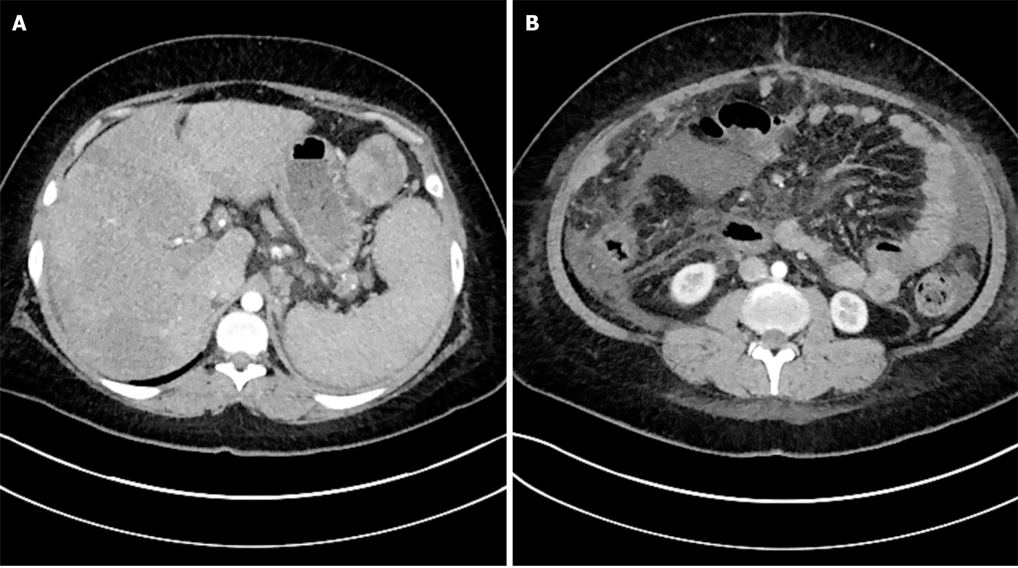
Figure 3 Computed tomography axial views of omental and peritoneal metastatic disease.
A: Computed tomography axial view showing left omental mass with liver metastases; B: Computed tomography axial view demonstrating peritoneal disease.
- Citation: Harris JT, Gurley S, Borazanci E. Adult pancreatoblastoma: Systematic review of the literature and case report of a young adult patient. World J Gastrointest Oncol 2025; 17(7): 106701
- URL: https://www.wjgnet.com/1948-5204/full/v17/i7/106701.htm
- DOI: https://dx.doi.org/10.4251/wjgo.v17.i7.106701


