Copyright
©The Author(s) 2025.
World J Gastrointest Oncol. Nov 15, 2025; 17(11): 112385
Published online Nov 15, 2025. doi: 10.4251/wjgo.v17.i11.112385
Published online Nov 15, 2025. doi: 10.4251/wjgo.v17.i11.112385
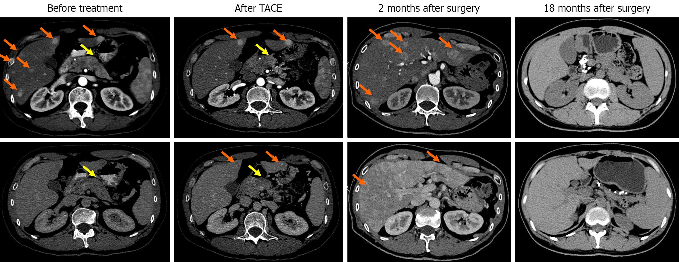
Figure 1 Computed tomography images of the patient before treatment, after transarterial chemoembolization, 2 months and 18 months after surgery.
The orange arrows point to the liver lesions, and the yellow arrows point to the pancreatic lesions. TACE: Transarterial chemoembolization.
Figure 2
The growth status of organoids from liver metastatic lesions in a pancreatic neuroendocrine tumor patient.
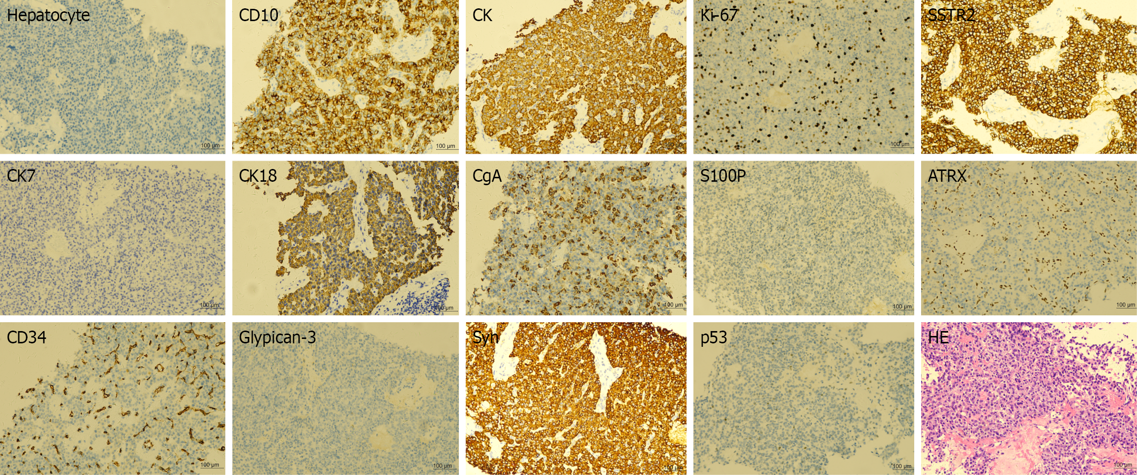
Figure 3 Immunohistochemical and hematoxylin and eosin staining results of the liver puncture tissue from a patient with pancreatic neuroendocrine tumors.
CK: Cytokeratin; CD: Cluster of differentiation; CgA: Chromogranin A; Syn: Synapsin; SSTR2: Somatostatin receptor 2; ATRX: X-linked alpha-thalassemia/mental retardation syndrome; HE: Hematoxylin and eosin.
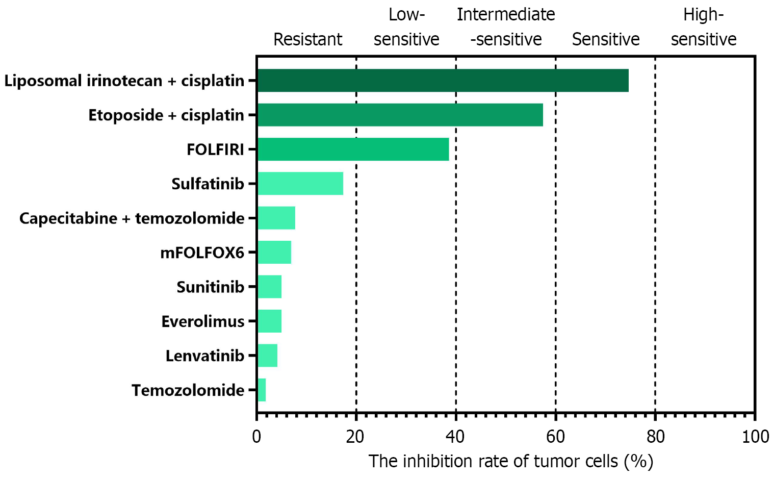
Figure 4 The growth status of organoids from liver metastatic lesions drug sensitivity testing result.
FOLFIRI: Folinic acid, fluorouracil, and irinotecan; mFOLFOX6: Modified folinic acid, fluorouracil, and oxaliplatin 6.
- Citation: Yang XM, Sun W, He YG, Peng XH, You N, Tang YC, Zheng L, Huang XB. Patient-derived organoids for the personalized treatment of pancreatic neuroendocrine tumor with liver metastases: A case report. World J Gastrointest Oncol 2025; 17(11): 112385
- URL: https://www.wjgnet.com/1948-5204/full/v17/i11/112385.htm
- DOI: https://dx.doi.org/10.4251/wjgo.v17.i11.112385


