©The Author(s) 2025.
World J Gastrointest Oncol. Jan 15, 2025; 17(1): 100210
Published online Jan 15, 2025. doi: 10.4251/wjgo.v17.i1.100210
Published online Jan 15, 2025. doi: 10.4251/wjgo.v17.i1.100210
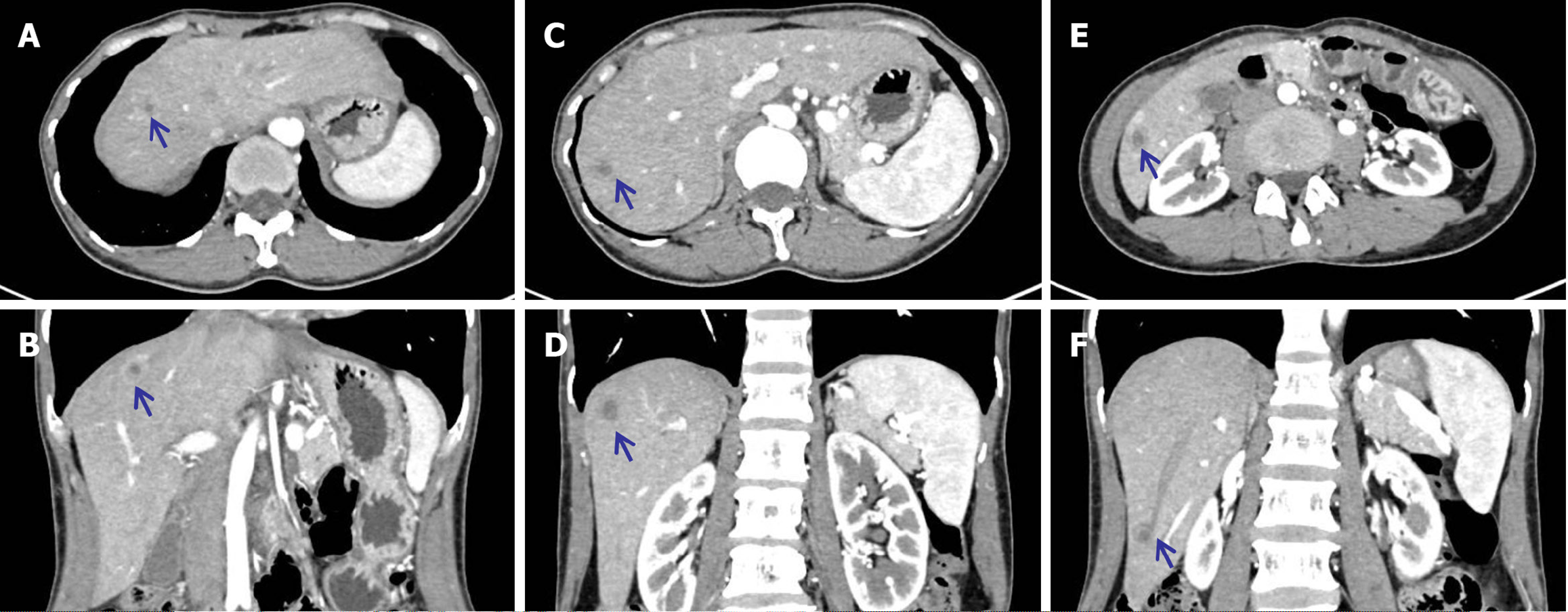
Figure 1 Baseline contrast-enhanced computed tomography revealed multiple liver nodules with a typical bull’s eye sign.
A and B: Central liver region; C and D: Subcapsular lesion; E and F: Inferior margin lesion. Blue arrow: Abnormal lesions in lateral and coronal sections of computed tomography.
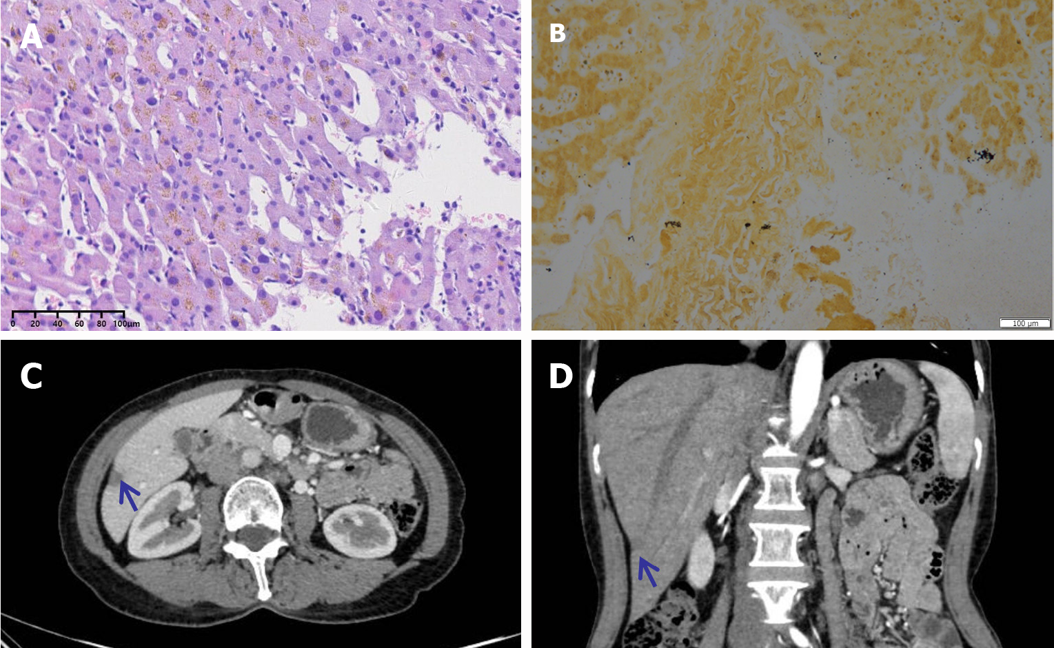
Figure 2 The patient’s final diagnosis, outcome, and follow-up.
A and B: Pathology results of hematoxylin-eosin and silver-impregnation staining showing hepatitis and Treponema pallidum; C and D: After penicillin G treatment, the number of nodules was significantly reduced, and they were well controlled. Blue arrow: Hepatic lesion in inferior margin after treatment.
- Citation: Wang YJ, Liu ZC, Wang J, Yang YM. Multiple liver metastases of unknown origin: A case report. World J Gastrointest Oncol 2025; 17(1): 100210
- URL: https://www.wjgnet.com/1948-5204/full/v17/i1/100210.htm
- DOI: https://dx.doi.org/10.4251/wjgo.v17.i1.100210


