©The Author(s) 2024.
World J Gastrointest Oncol. Aug 15, 2024; 16(8): 3624-3634
Published online Aug 15, 2024. doi: 10.4251/wjgo.v16.i8.3624
Published online Aug 15, 2024. doi: 10.4251/wjgo.v16.i8.3624
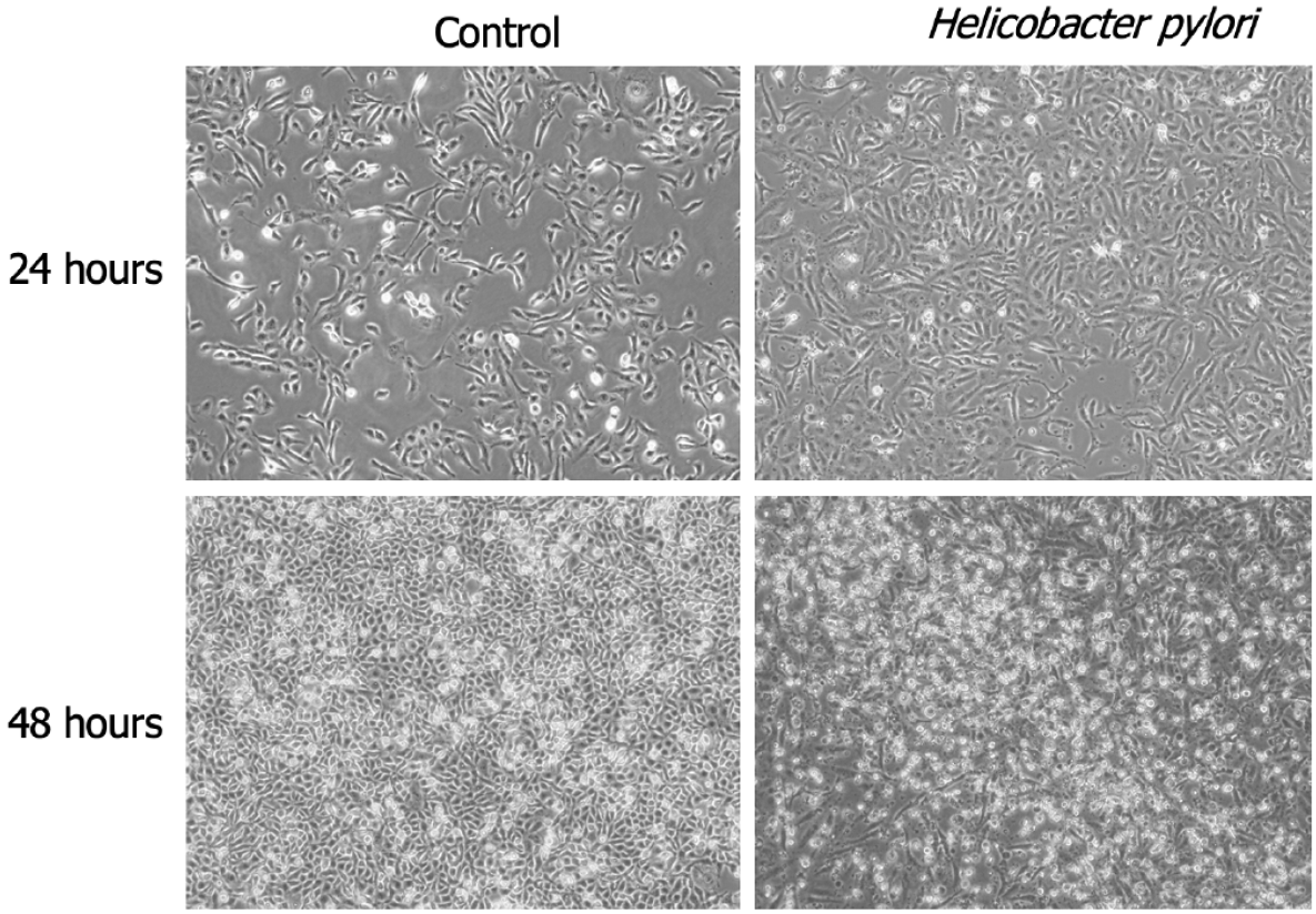
Figure 1 Significant morphological alterations in GES-1 cells following exposure to Helicobacter pylori for 24 h and 48 h.
A: 24 h; B: 48 h. × 40 magnification.
Figure 2 Helicobacter pylori infection reduces the viability of GES-1 gastric epithelial cells.
A and B: Stimulation of cells for 24 h or 48 h at specified ratios of cells to Helicobacter pylori (H. pylori); C: Viability of GES-1 cells treated with different ratios of H. pylori over various time periods. The viability of the control group (GES-1 cells not stimulated with H. pylori) was set at 100%. Significant differences were observed: aP < 0.05, bP < 0.01, cP < 0.001, dP < 0.0001 vs control group.
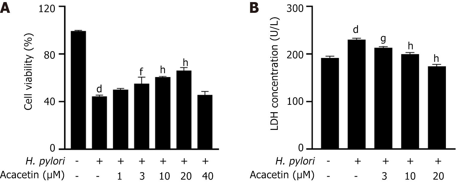
Figure 3 Acacetin inhibits the decline in cell viability and lactate dehydrogenase release induced by Helicobacter pylori infection.
A: Cells were pre-treated with specified concentrations of acacetin extract for 2 h, followed by stimulation with Helicobacter pylori (H. pylori) [multiplicity of infection (MOI) = 1:100] for 24 h; B: After pretreatment with specified concentrations of acacetin extract for 2 h and subsequent stimulation with H. pylori (MOI = 1:100) for 24 h, the amount of lactate dehydrogenase released into the cell culture supernatant was measured. aP < 0.05, bP < 0.01, cP < 0.001, dP < 0.0001 vs control group; eP < 0.05, fP < 0.01, gP < 0.001, hP < 0.0001 vs H. pylori group.
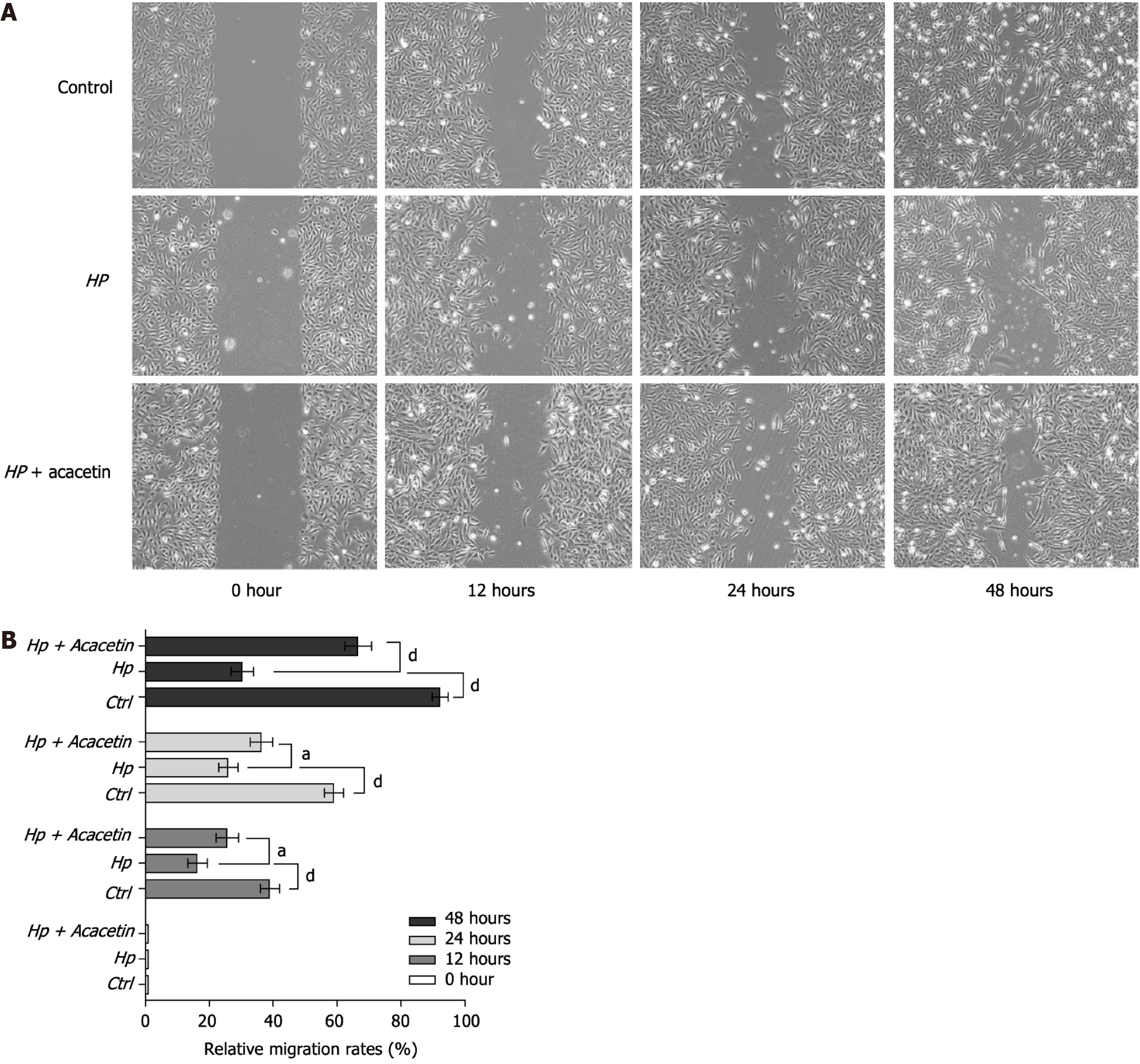
Figure 4 Migration analysis was performed through a wound healing assay.
A: Images of the wound region captured at four distinct time intervals (0, 12, 24, and 48 h). × 40 magnification; B: The area of wound closure was analyzed and converted into migration rates using ImageJ software. HP: Helicobacter pylori.
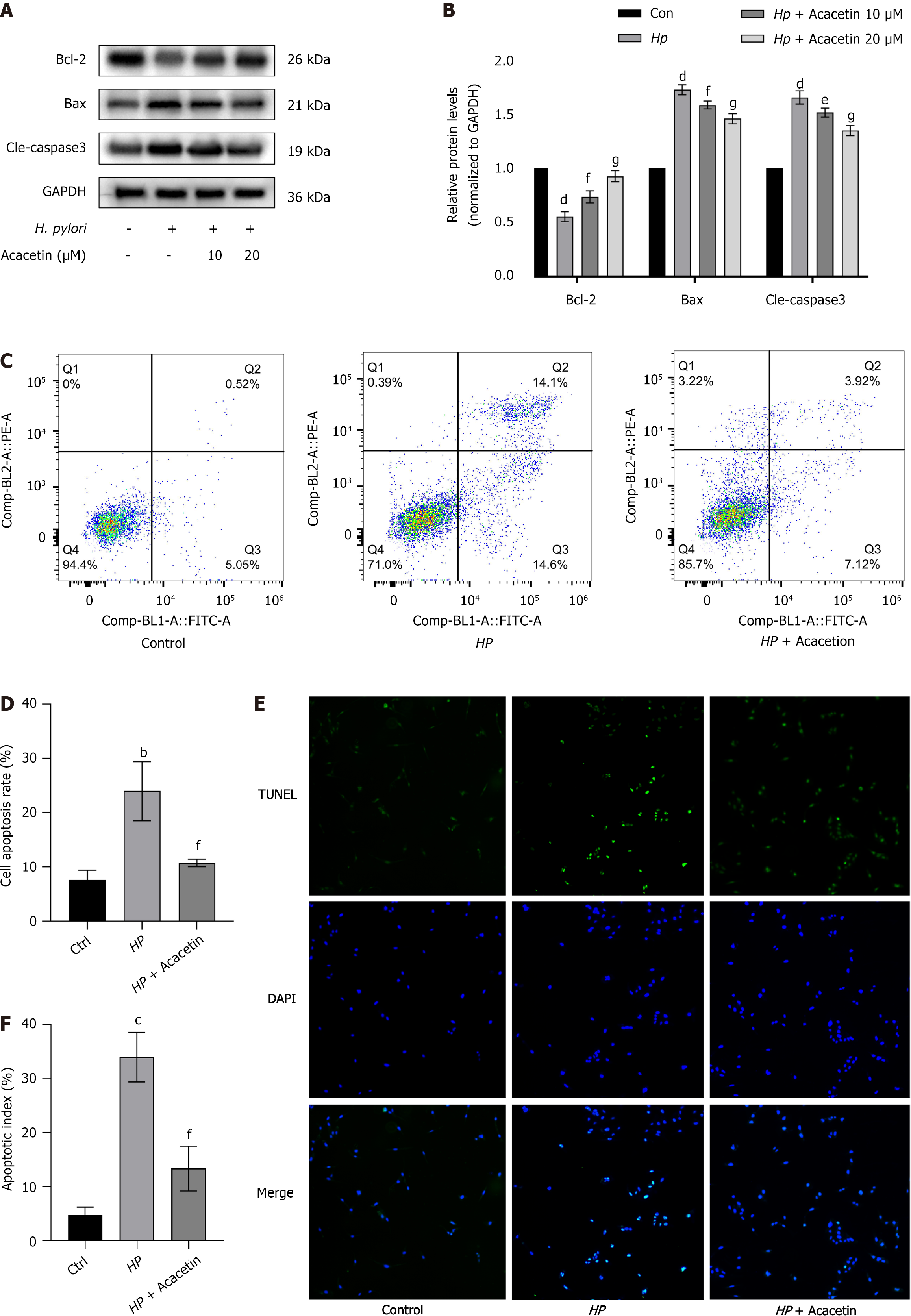
Figure 5 Acacetin extract inhibits Helicobacter pylori-induced apoptosis in GES-1 cells.
A and B: Protein levels of cleaved caspase-3, Bcl-2, and Bax were analyzed by immunoblotting following pre-treatment with acacia extract (10 µmol/L, 20 µmol/L) and subsequent stimulation with Helicobacter pylori(H. pylori) (cell to H. pylori ratio of 1:100) for 24 h. GAPDH served as an internal control, with the ratio in the blank control group set to 1; C and D: Apoptosis levels in the blank group, H. pylori-infected group, and H. pylori plus acacetin extract group were measured by flow cytometry; E: Representative images of cells stained with TUNEL and DAPI. × 40 magnification; F: Percentage of apoptotic cells quantified as the apoptosis index.
- Citation: Yao QX, Li ZY, Kang HL, He X, Kang M. Effect of acacetin on inhibition of apoptosis in Helicobacter pylori-infected gastric epithelial cell line. World J Gastrointest Oncol 2024; 16(8): 3624-3634
- URL: https://www.wjgnet.com/1948-5204/full/v16/i8/3624.htm
- DOI: https://dx.doi.org/10.4251/wjgo.v16.i8.3624


