Copyright
©The Author(s) 2024.
World J Gastrointest Oncol. Mar 15, 2024; 16(3): 979-990
Published online Mar 15, 2024. doi: 10.4251/wjgo.v16.i3.979
Published online Mar 15, 2024. doi: 10.4251/wjgo.v16.i3.979
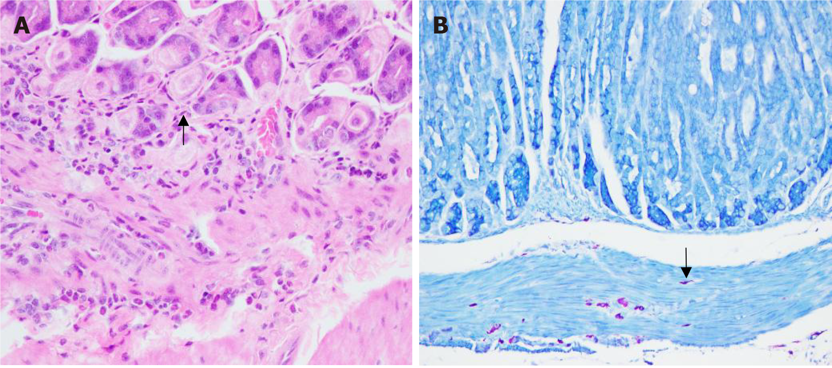
Figure 1 Staining results of mouse gastric sinus tissue.
A: HE staining of mouse gastric antrum tissue (400 ×); B: Gimesa-stained image of mouse gastric sinus tissue (200 ×). The arrows indicate the location of the Helicobacter pylori.
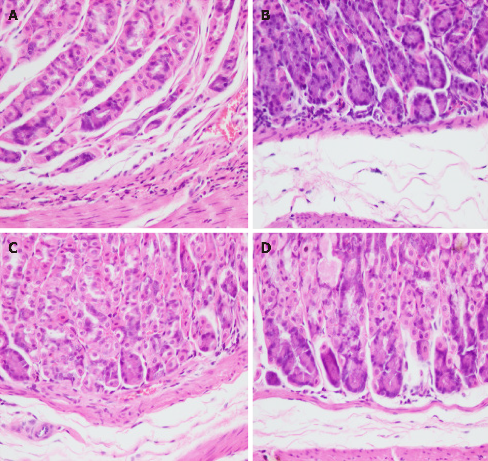
Figure 2 Pathological observation of HE stain of mouse stomach tissue (400 ×).
A: Control group; B: N-methyl-N-nitrosourea (MNU) group; C: Helicobacter pylori + MNU group; D: Moluodan group.
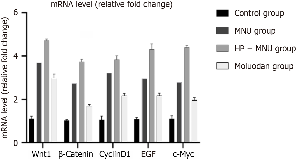
Figure 3 Relative expression of Wnt1, β-catenin, cyclinD1, epidermal growth factor, c-Myc m-RNA in each group.
EGF: Epidermal growth factor; MNU: N-methyl-N-nitrosourea; H. pylori: Helicobacter pylori.
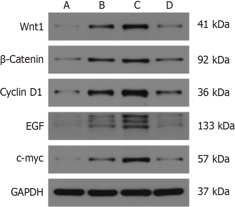
Figure 4 Western blot of Wnt1, β-catenin, cyclinD1, epidermal growth factor, and c-Myc protein expression in each group.
A: Control group; B: Helicobacter pylori +N-methyl-N-nitrosourea (MNU) group; C: MNU group; D: Moluodan group.
- Citation: Wang YM, Luo ZW, Shu YL, Zhou X, Wang LQ, Liang CH, Wu CQ, Li CP. Effects of Helicobacter pylori and Moluodan on the Wnt/β-catenin signaling pathway in mice with precancerous gastric cancer lesions. World J Gastrointest Oncol 2024; 16(3): 979-990
- URL: https://www.wjgnet.com/1948-5204/full/v16/i3/979.htm
- DOI: https://dx.doi.org/10.4251/wjgo.v16.i3.979


