Copyright
©The Author(s) 2024.
World J Gastrointest Oncol. Mar 15, 2024; 16(3): 699-715
Published online Mar 15, 2024. doi: 10.4251/wjgo.v16.i3.699
Published online Mar 15, 2024. doi: 10.4251/wjgo.v16.i3.699
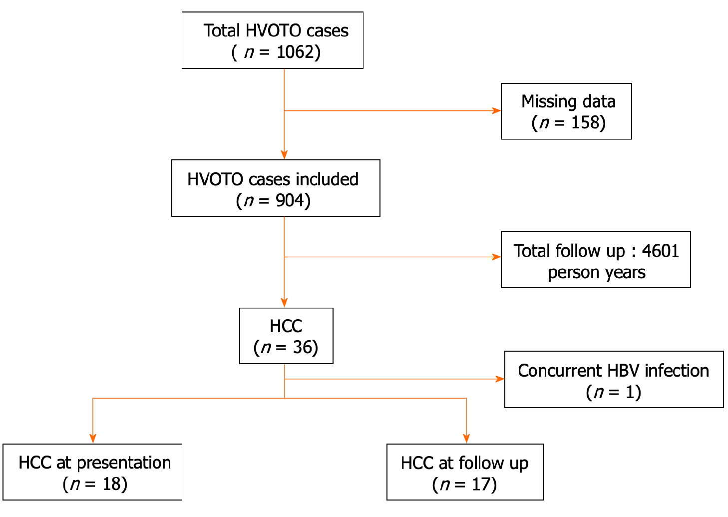
Figure 1 Risk of hepatocellular carcinoma in patients with Budd-Chiari syndrome.
HBV: Hepatitis B virus; HCC: Hepatocellular carcinoma; HVOTO: Hepatic venous outflow tract obstruction.
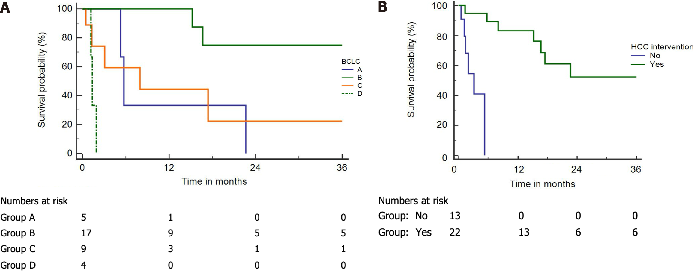
Figure 2 Kaplan-Meier curves of Budd-Chiari syndrome-hepatocellular carcinoma patients.
A and B: Kaplan-Meier curves of Budd-Chiari syndrome-hepatocellular carcinoma (HCC) patients; A: Per the Barcelona Clinic Liver Cancer (BCLC) stages (log-rank P < 0.001); B: Intervention and without intervention (log-rank P < 0.001).
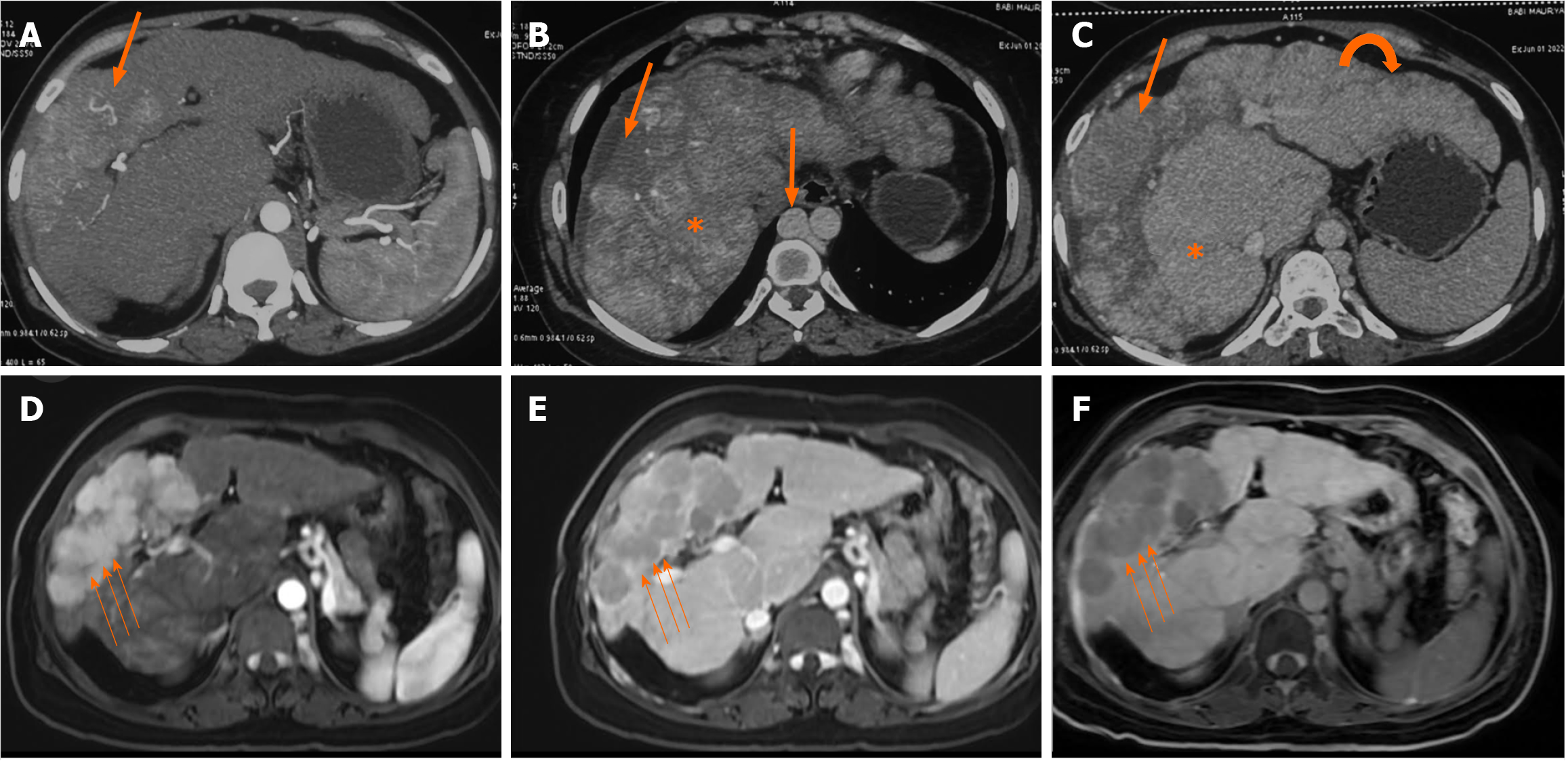
Figure 3 Axial multiphase computed tomography images.
A-C: Axial multiphase computed tomography images showed a large arterial phase enhancing lesion (arrow) in the arterial phase and washout in portovenous phase (B) and delayed phase (C) in segment VIII and IV of the liver with background liver showing features of congestive changes (asterix) and cirrhotic changes (curved arrow). Note: Dilated azygous system due to inferior vena cava obstruction (block arrow); D and E: Axial multiphase magnetic resonance imaging (post angioplasty) showed resolution of congestive changes and normal caliber azygous system; F: Non-retention of contrast in hepatobiliary phase. Large arterial phase enhancing lesion (small arrows) in arterial phase (A) with washout in portovenous phase (E) and non-retention of contrast in hepatobiliary phase (F).
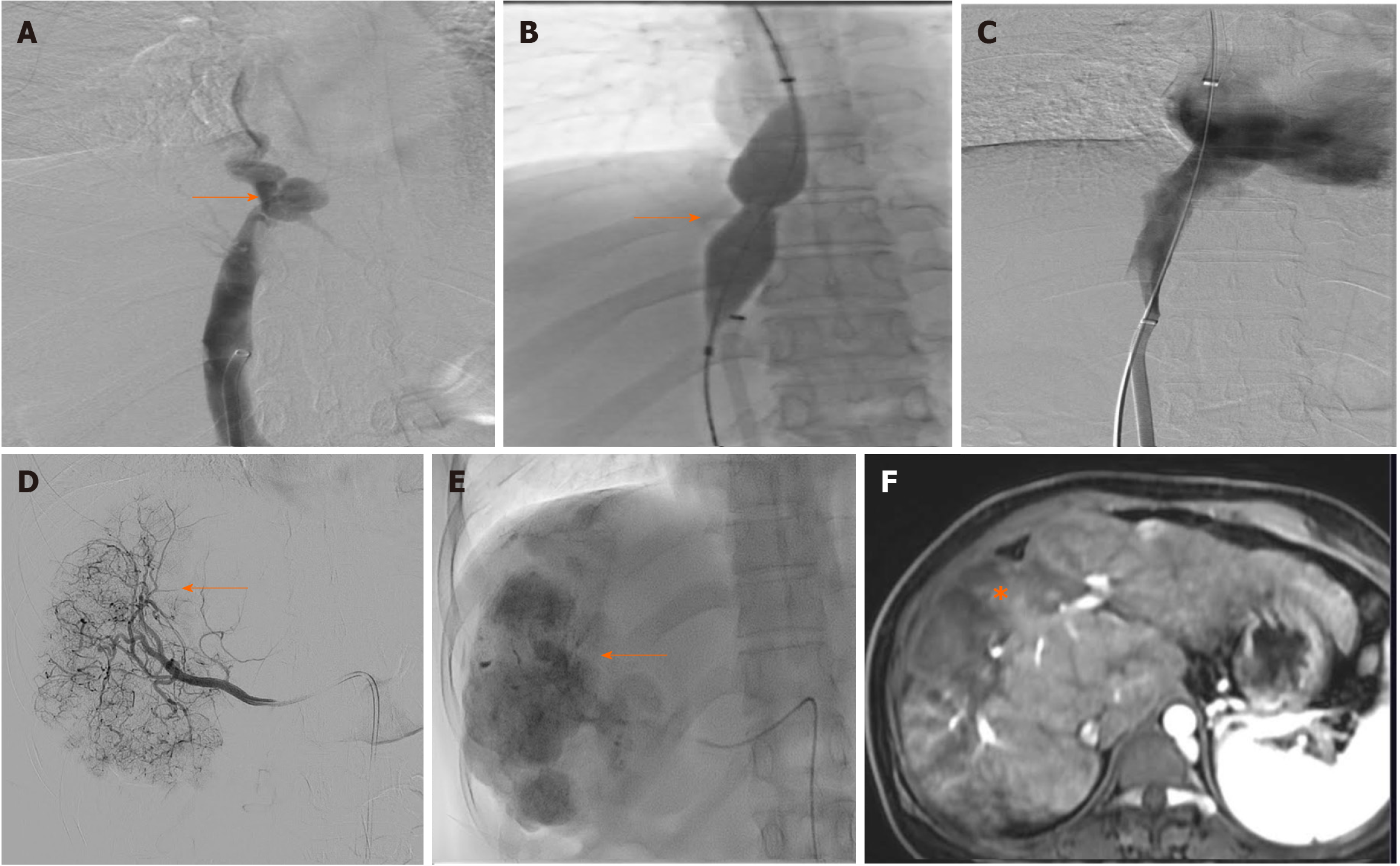
Figure 4 Digital subtraction spot images.
A-F: Digital subtraction spot images showed short segment narrowing of inferior vena cava (A, arrow) which was dilated using 20 mm × 40 mm balloon catheter (B, arrow), post angioplasty angiogram (C) good flow across the inferior vena cava without any residual narrowing. Selective right hepatic angiogram showed tumor blush (D, arrow), which was treated using lipiodol transarterial chemoembolization (E, arrow), follow-up magnetic resonance imaging after transarterial chemoembolization showed no residual enhancing lesion in the treated lesion (F, asterisk).
- Citation: Agarwal A, Biswas S, Swaroop S, Aggarwal A, Agarwal A, Jain G, Elhence A, Vaidya A, Gupte A, Mohanka R, Kumar R, Mishra AK, Gamanagatti S, Paul SB, Acharya SK, Shukla A, Shalimar. Clinical profile and outcomes of hepatocellular carcinoma in primary Budd-Chiari syndrome. World J Gastrointest Oncol 2024; 16(3): 699-715
- URL: https://www.wjgnet.com/1948-5204/full/v16/i3/699.htm
- DOI: https://dx.doi.org/10.4251/wjgo.v16.i3.699


