Copyright
©The Author(s) 2024.
World J Gastrointest Oncol. Mar 15, 2024; 16(3): 614-629
Published online Mar 15, 2024. doi: 10.4251/wjgo.v16.i3.614
Published online Mar 15, 2024. doi: 10.4251/wjgo.v16.i3.614
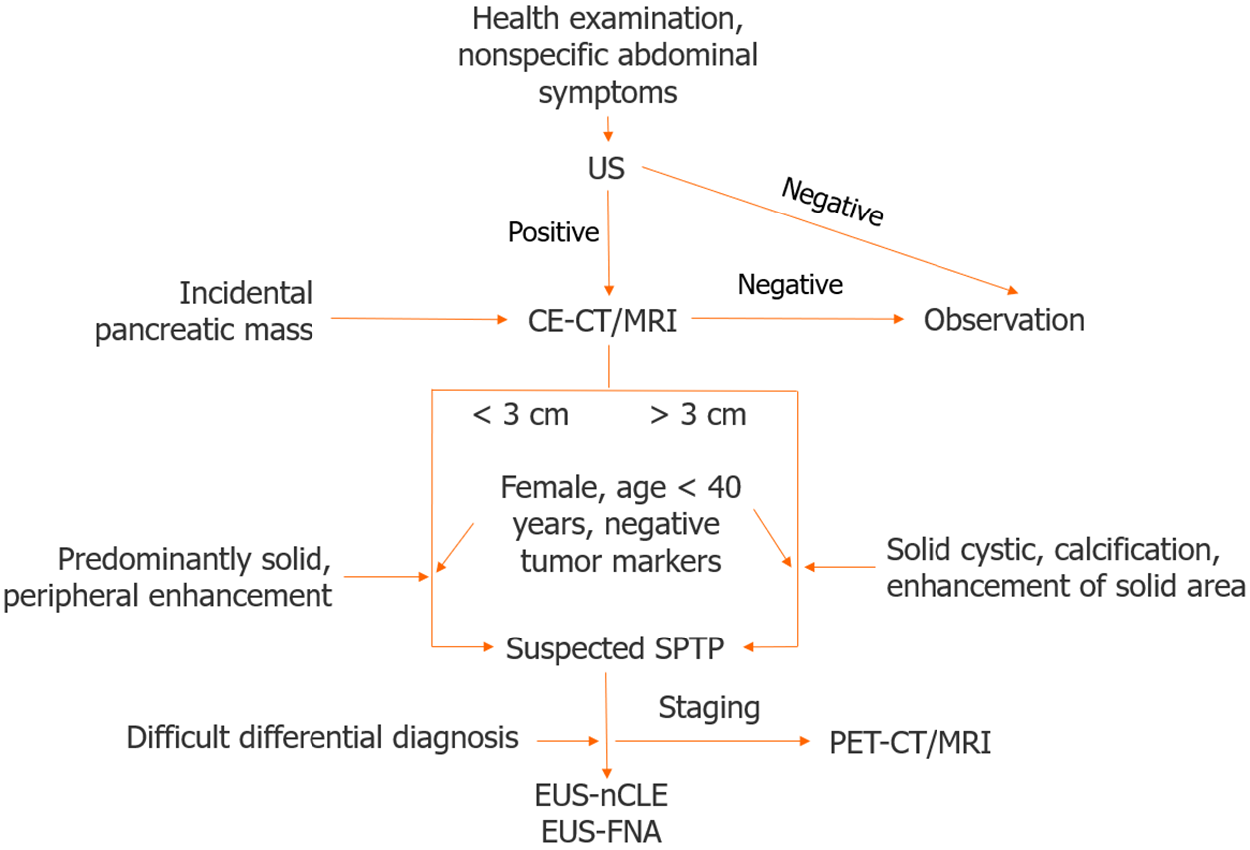
Figure 1 Diagnosis algorithm for solid pseudopapillary tumor of the pancreas.
US: Ultrasound; CE-CT: Contrast enhanced computed tomography; MRI: Magnetic resonance imaging; SPTP: Solid pseudopapillary tumor of the pancreas; EUS-FNA: Endoscopic ultrasound guided fine-needle aspiration; EUS-nCLE: Endoscopic ultrasound guided needle-based confocal laser endomicroscopy; PET-CT: Positron emission tomography-computed tomography; PET-MRI: Positron emission tomography-magnetic resonance imaging.
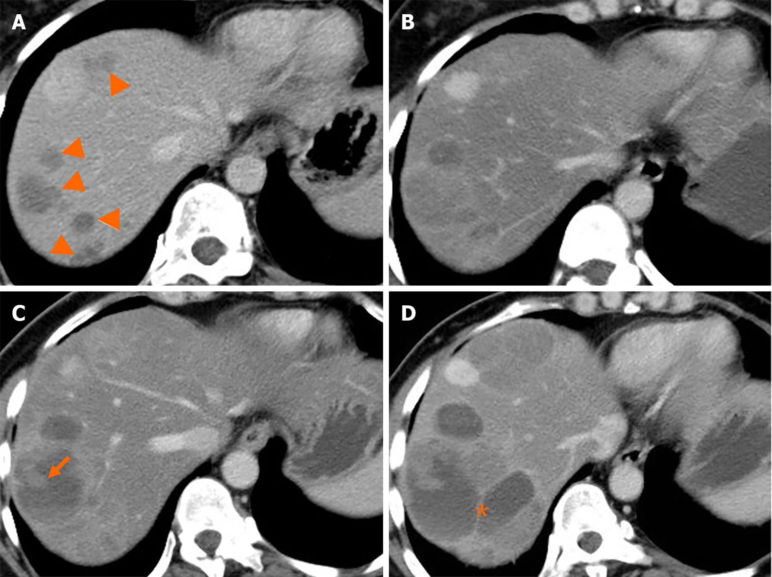
Figure 2 Natural history (classical type) of liver metastasis from solid pseudopapillary tumor of the pancreas.
A and B: Multiple liver metastases (A, arrowheads) were detected 38 months after surgery. Five months later, the lesions increased in size gradually (B); C and D: Cystic change of the lesion with solid component (C, arrow) was shown after 10 months. Seventeen months later, the lesions continued increasing with septum-like structure detected (D, asterisk).
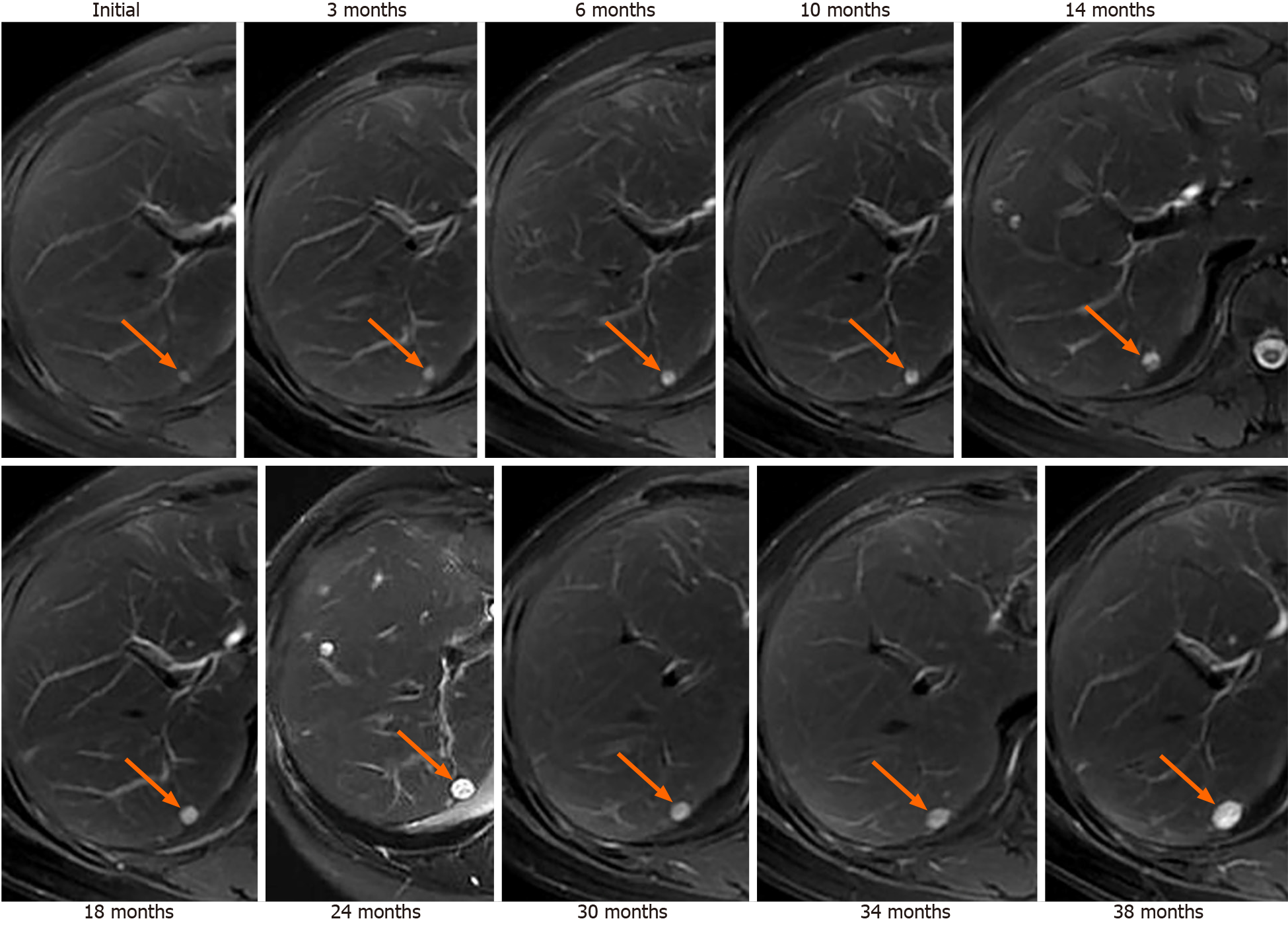
Figure 3 Indolent type of liver metastasis from solid pseudopapillary tumor of the pancreas.
A 46-year-old woman, who had a history of surgery for malignant solid pseudopapillary tumor of the pancreas with Ki-67 of 1%, received serial magnetic resonance imaging scans as surveillance. T2WI showed a new-onset homogeneous high signal nodule in the liver segment 7 (arrows). During the 3-year follow-up, the lesion grew slightly.
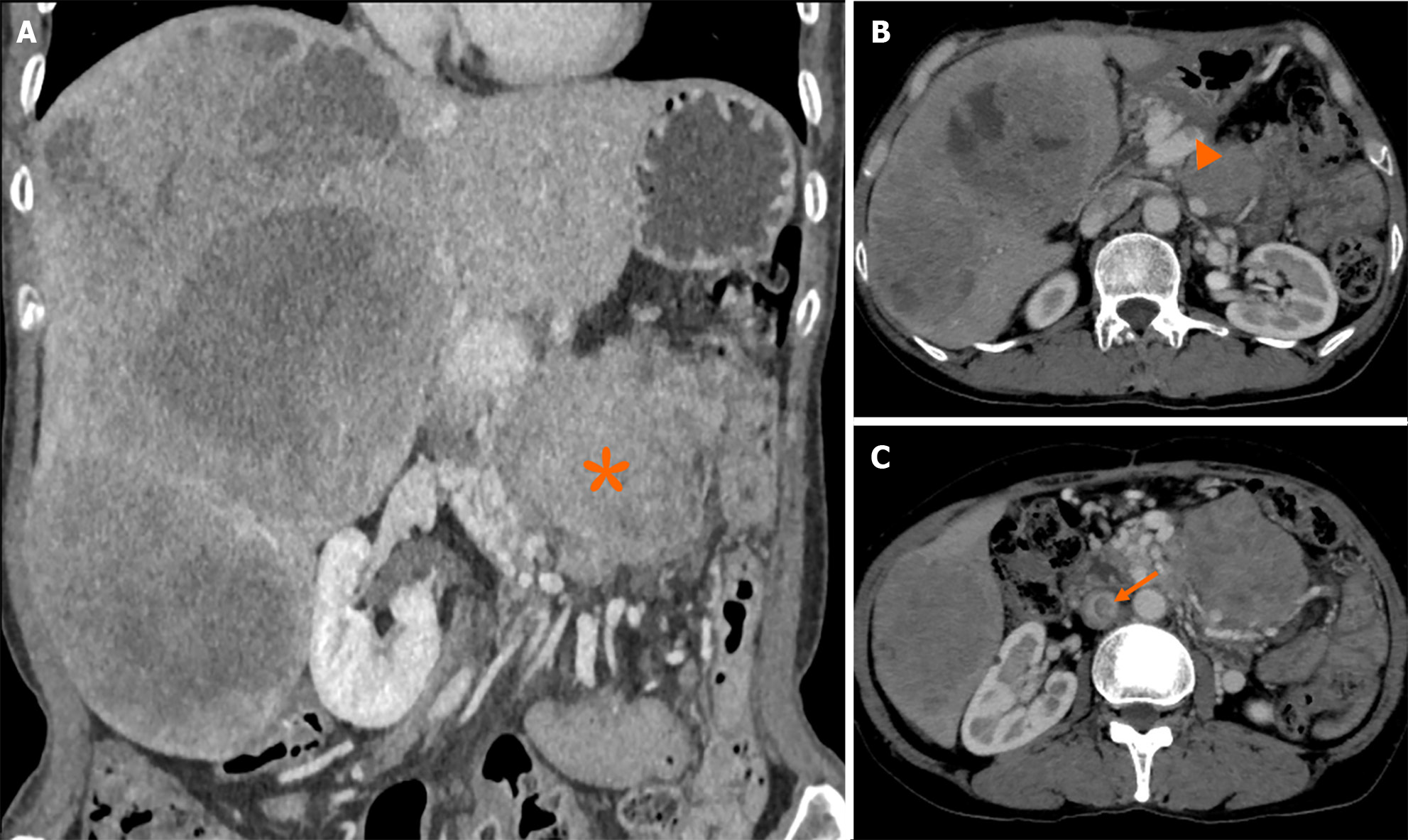
Figure 4 Aggressive type of liver metastasis from solid pseudopapillary tumor of the pancreas.
A 55-year-old woman, who underwent surgery for solid pseudopapillary tumor of the pancreas 6 years ago, presented with incidental detection of abdominal masses after abdominal blunt trauma due to an accidental fall. Computed tomography scan showed multiple heterogeneously enhancing masses with extensive central necrosis fused in the right liver, with a size of 26 cm × 17 cm. A-C: A retroperitoneal mass (A, asterisk) invading the superior mesenteric vein (SMV) was detected, with presence of collateral circulation, and tumor thrombi filling the SMV (B, arrowheads) and inferior vena cava (C, arrow).
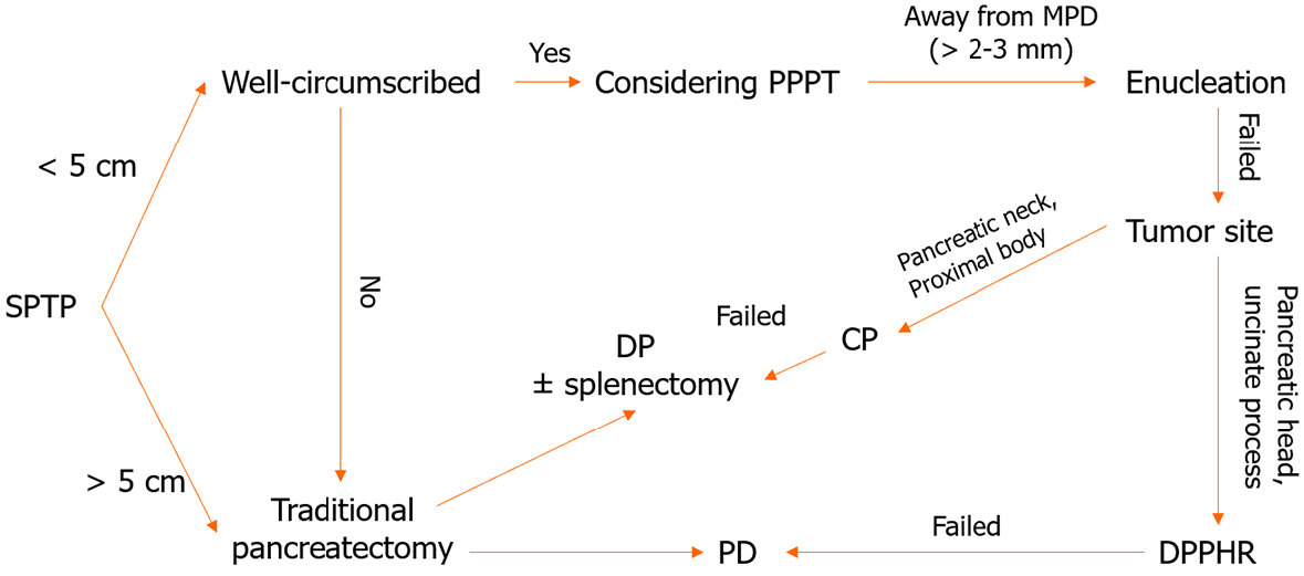
Figure 5 The algorithm for surgical treatment of solid pseudopapillary tumor of the pancreas.
SPTP: Solid pseudopapillary tumor of the pancreas; PPPT: Parenchyma-preserving pancreatectomy; MPD: Main pancreatic duct; CP: Central pancreatectomy; DP: Distal pancreatectomy; PD: Pancrea
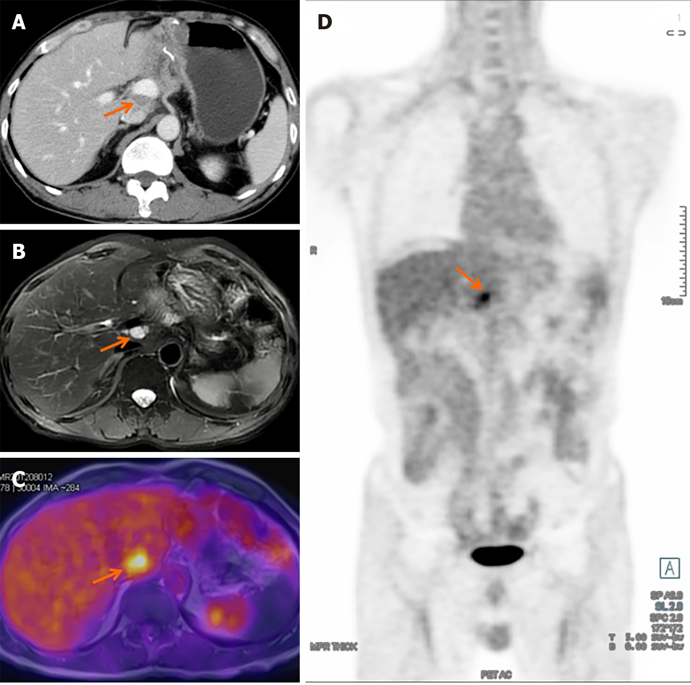
Figure 6 Detection of recurrence in solid pseudopapillary tumor of the pancreas.
A: Abdominal enhanced computed tomography scan revealed a hypodense mass between the portal vein and inferior vena cava; B: T2WI magnetic resonance imaging (MRI) demonstrated a high signal mass located at the hepatic hilum; C and D: Axial-fused (C) and coronal-fused (D) positron emission tomography/MRI showed that the lesion had increased fluorodeoxyglucose uptake.
- Citation: Xu YC, Fu DL, Yang F. Unraveling the enigma: A comprehensive review of solid pseudopapillary tumor of the pancreas. World J Gastrointest Oncol 2024; 16(3): 614-629
- URL: https://www.wjgnet.com/1948-5204/full/v16/i3/614.htm
- DOI: https://dx.doi.org/10.4251/wjgo.v16.i3.614


