©The Author(s) 2024.
World J Gastrointest Oncol. Dec 15, 2024; 16(12): 4746-4752
Published online Dec 15, 2024. doi: 10.4251/wjgo.v16.i12.4746
Published online Dec 15, 2024. doi: 10.4251/wjgo.v16.i12.4746
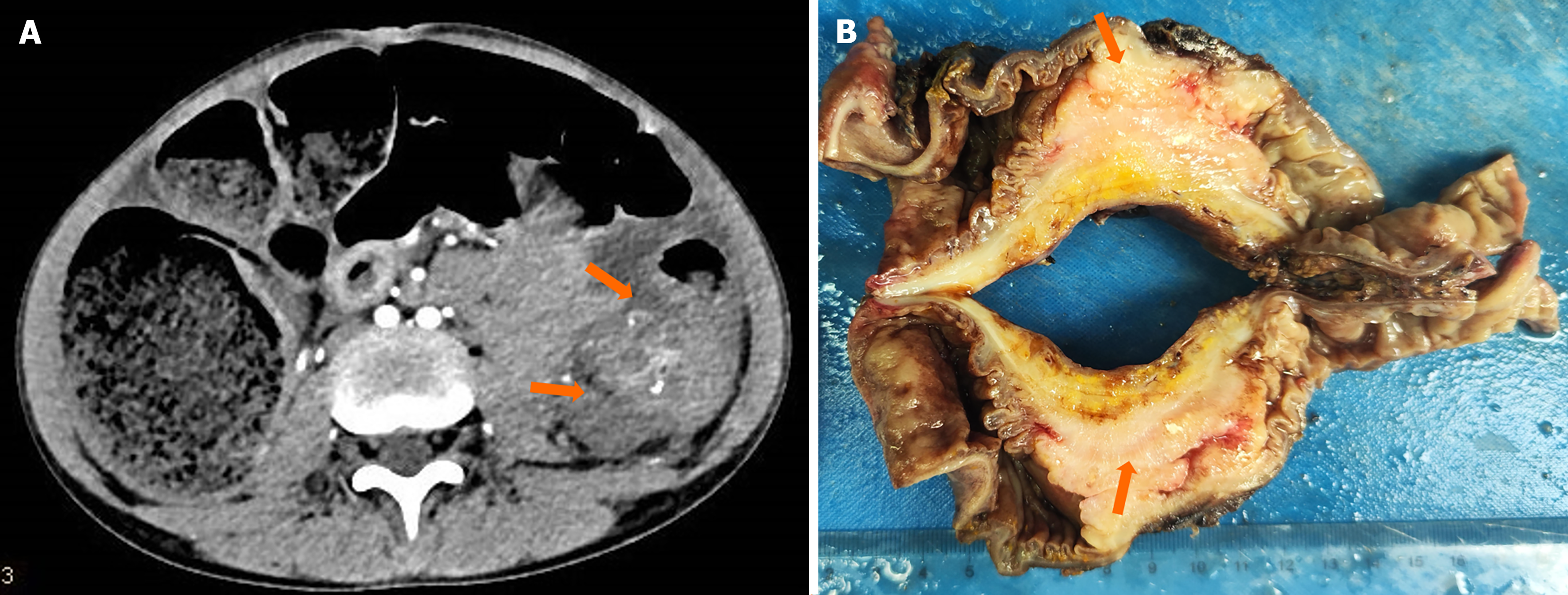
Figure 1 Tumor imaging data and surgical gross specimens.
A: Computed tomography scan revealed irregular masses of soft tissue density in the transverse colon, exhibiting heterogeneous density and multiple calcifications. The lesion exerted pressure on the adjacent descending colon, resulting in obstruction of the proximal transverse colon (orange arrowhead); B: Gross specimen of the tumor (orange arrowhead).
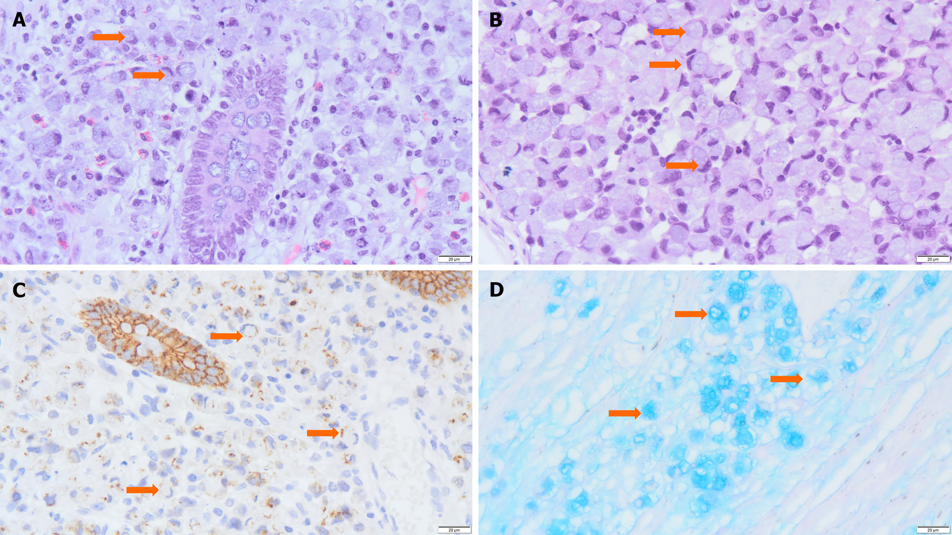
Figure 2 Pathological features of signet-ring cell carcinoma.
A: Tumor cells were distributed diffusely around the normal mucosal glands. Most tumor cells exhibited a signet-ring appearance due to cytoplasmic mucus crimping their nuclei (orange arrowhead) [hematoxylin and eosin (H&E), 400 ×]; B: Typical signet ring cells (orange arrowhead) (H&E, 400 ×); C: E-cadherin (+) showing E-cadherin was weaker in tumor cells with poor adhesion (orange arrowhead) (immunohistochemistry, 400 ×); D: Alcian Blue/Phosphoric Acid Schiff staining revealed blue-stained mucus distributed both inside and outside the cells (orange arrowhead indicates mucus) (special staining, 400 ×).
- Citation: Lv L, Song YH, Gao Y, Pu SQ, A ZX, Wu HF, Zhou J, Xie YC. Signet-ring cell carcinoma of the transverse colon in a 10-year-old girl: A case report. World J Gastrointest Oncol 2024; 16(12): 4746-4752
- URL: https://www.wjgnet.com/1948-5204/full/v16/i12/4746.htm
- DOI: https://dx.doi.org/10.4251/wjgo.v16.i12.4746


