Copyright
©The Author(s) 2023.
World J Gastrointest Oncol. Dec 15, 2023; 15(12): 2120-2137
Published online Dec 15, 2023. doi: 10.4251/wjgo.v15.i12.2120
Published online Dec 15, 2023. doi: 10.4251/wjgo.v15.i12.2120
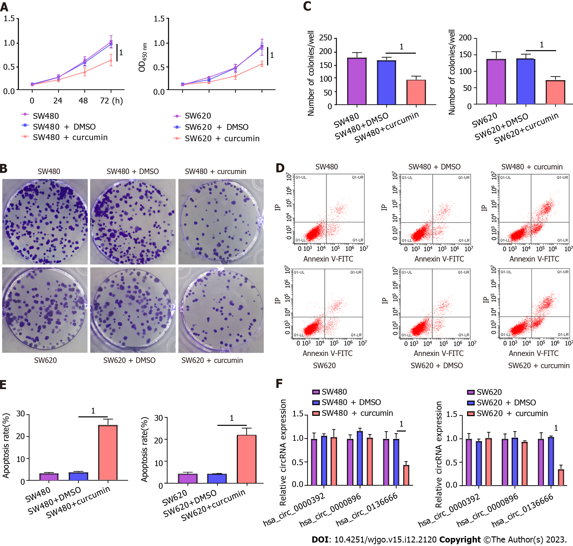
Figure 1 The impacts of curcumin treatment on colorectal carcinoma (SW480 and SW620) cell proliferation and apoptosis.
Colorectal carcinoma (CRC) cells were treated with DMSO or curcumin (20 μM). A: Cell viability was detected in treated or untreated SW480 and SW620 using cell counting kit-8 assays; B and C: Colony number assessment by colony-forming cell assays in treated or untreated SW480 and SW620; D and E: Apoptosis rate analysis in treated or untreated CRC cells by flow cytometry assays; F: Real-time quantitative PCR analysis of hsa_circ_0000392, hsa_circ_0000896, and hsa_circ_0136666 expression in treated or untreated SW480 and SW620 cells. 1P < 0.05.
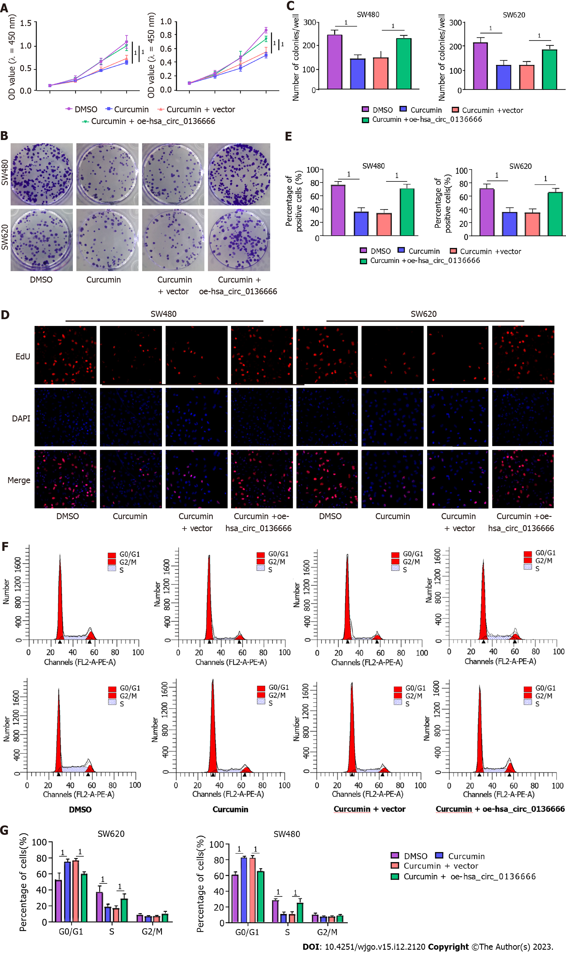
Figure 2 Overexpressing hsa_circ_0136666 overturned the effect of curcumin on proliferation in colorectal carcinoma SW480 and SW620 cells.
Colorectal carcinoma cells were treated with DMSO, curcumin, curcumin + vector, and curcumin + oe-hsa_circ_0136666. A: Cell counting kit-8 assays examined SW480 and SW620 cell viability after treatment; B and C: Colony-forming cell assays detected colony numbers in treated SW480 and SW620; D and E: 5-Ethynyl-2’-deoxyuridine assays measured positive cells in treated SW480 and SW620; F and G: Flow cytometry assays analyzed cell cycle distribution in treated SW480 and SW620. 1P < 0.05.
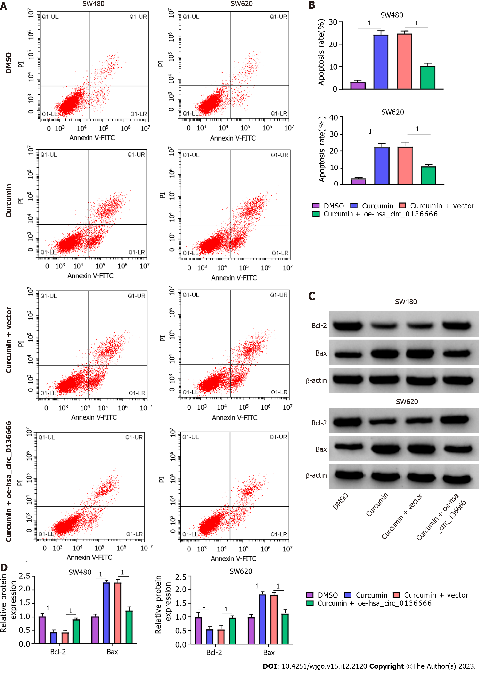
Figure 3 Upregulating hsa_circ_0136666 overturned curcumin-mediated apoptosis promotion in colorectal carcinoma cells.
SW480 and SW620 were treated with DMSO, curcumin, curcumin + vector, and curcumin + oe-hsa_circ_0136666. A and B: Flow cytometry assays tested apoptosis rate in treated SW480 and SW620; C and D: Western blot assays determined Bcl-2 and Bax protein levels in treated SW480 and SW620. 1P < 0.05.
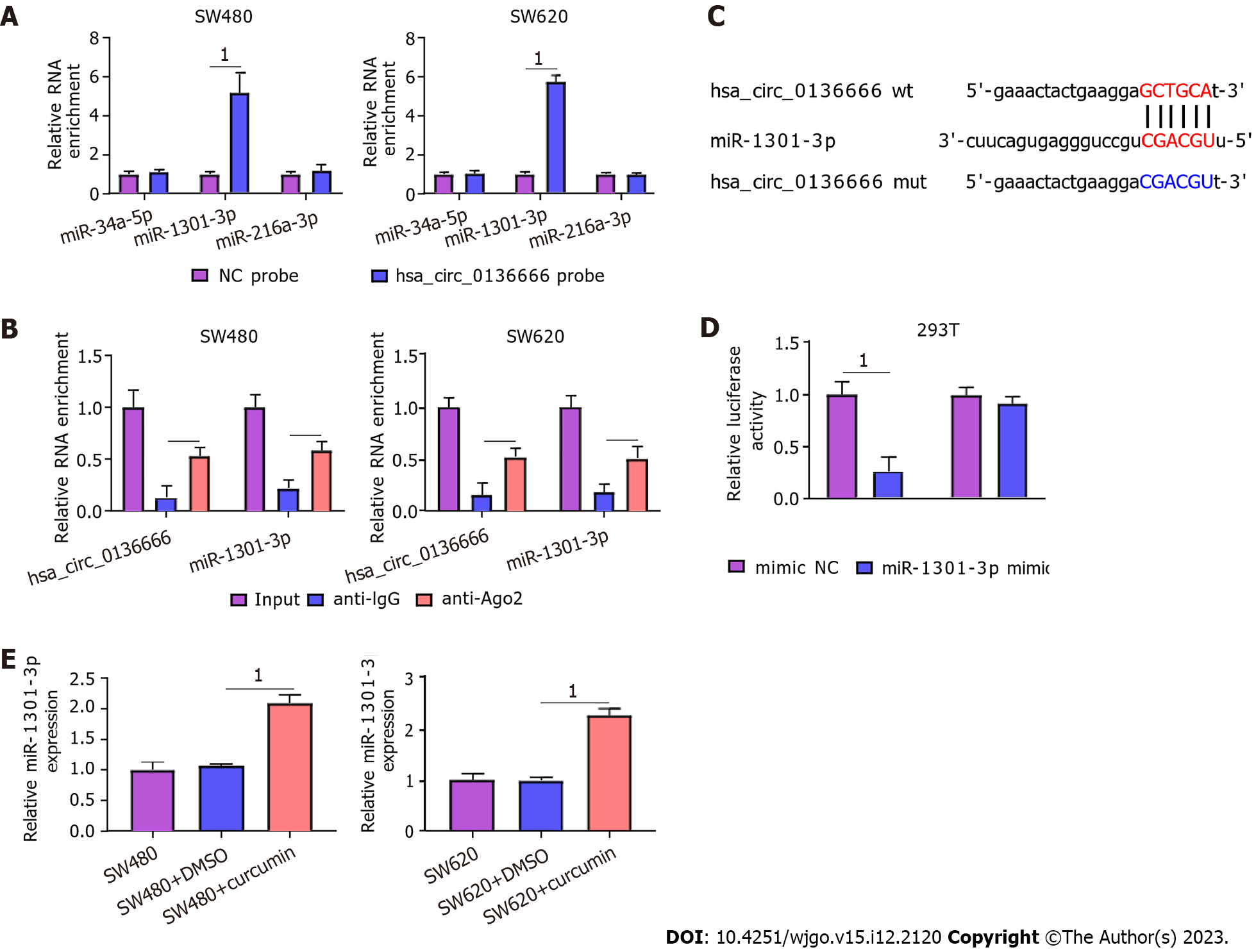
Figure 4 Hsa_circ_0136666 directly bound to miR-1301-3p.
A: Real-time quantitative PCR (RT-qPCR) measured relative levels of 3 miRNA candidates in SW480 and SW620 lysates; B: RNA immunoprecipitation assays assessed miR-1301-3p endogenously associated with hsa_circ_0136666 in SW480 and SW620 extracts; C: Binding loci between hsa_circ_0136666 and miR-1301-3p and the hsa_circ_0136666 mut sequence; D: The binding relationship was verified by a dual-luciferase reporter assay; E: RT-qPCR assays quantified miR-1301-3p in SW480, SW480 + DMSO, SW480 + curcumin, SW620, SW620 + DMSO, and SW620 + curcumin. 1P < 0.05. wt: Wild-type; mut: Mutant-type.
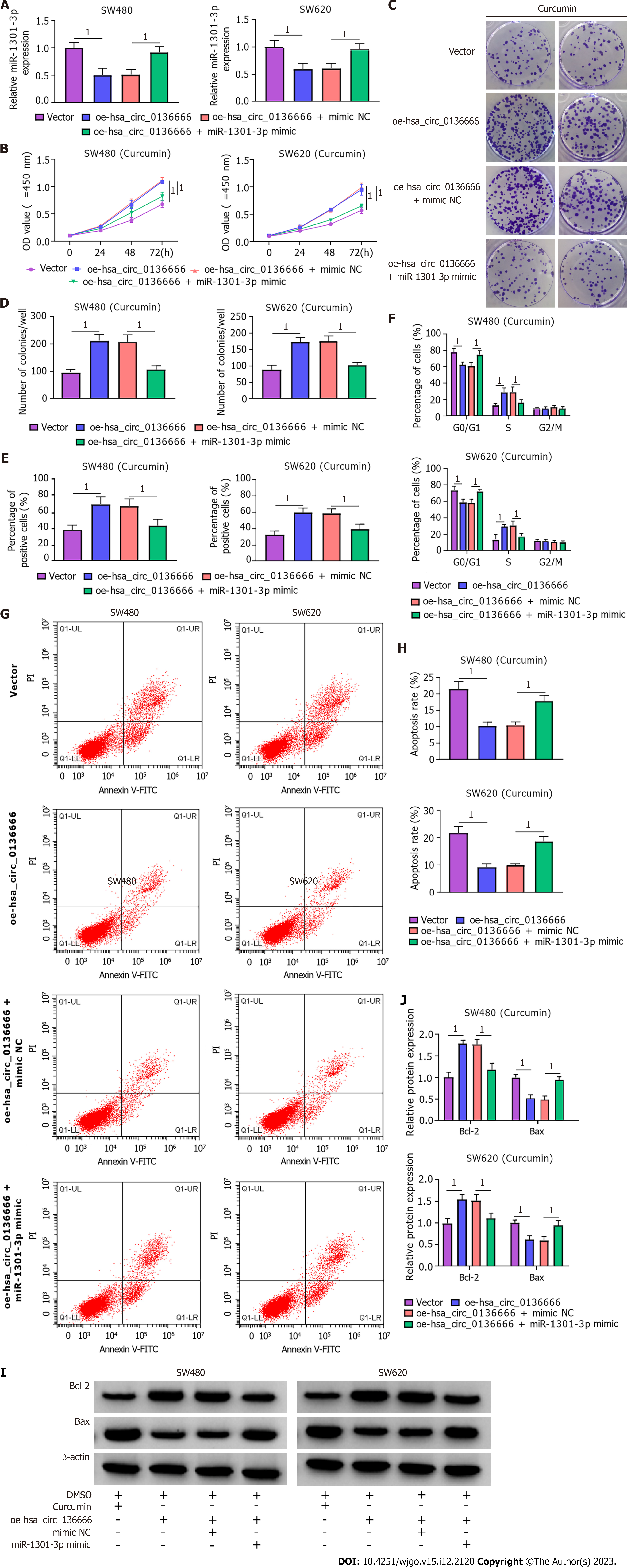
Figure 5 miR-1301-3p overturned the effect of hsa_circ_0136666 on growth and apoptosis in curcumin-treated colorectal carcinoma cells.
A: Real-time quantitative PCR assays quantified miR-1301-3p levels in SW480 and SW620 transfected with vector, oe-hsa_circ_0136666, oe-hsa_circ_0136666+mimic NC, and oe-hsa_circ_0136666 + miR-1301-3p mimic; B-J: Colorectal carcinoma cells were intervened by curcumin after transfection; B: Cell counting kit-8 assays measured cell viability; C and D: Colony-forming cell assays determined the colony number; E: 5-Ethynyl-2’-deoxyuridine assays examined positive cell count; F: Flow cytometry assays determined cell cycle distribution; G and H: Flow cytometry assays tested apoptosis; I and J: Western blot assays quantified Bcl-2 and Bax protein levels. 1P < 0.05.
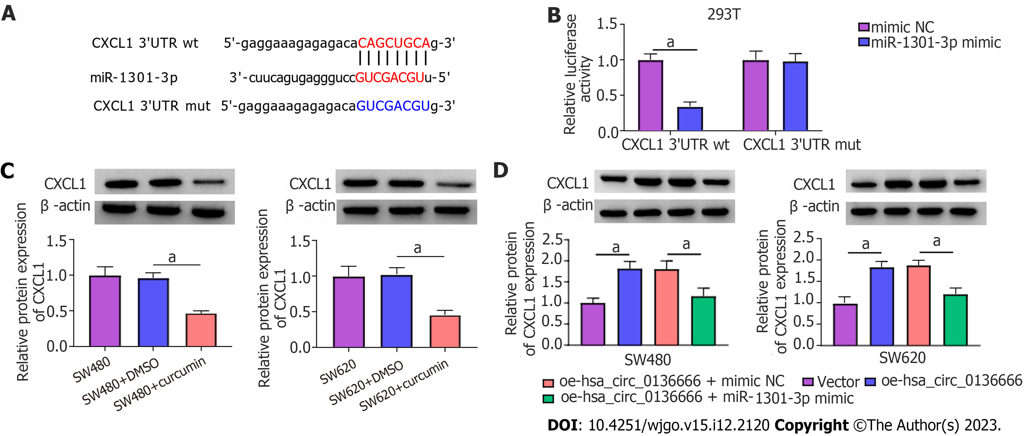
Figure 6 CXCL1 was miR-1301-3p’s direct target.
A: Putative binding sequences between miR-1301-3p and CXCL1 3’UTR, and mutant sites in CXCL1 3’UTR mut; B: Validation of prediction by dual-luciferase reporter assay; C: Western blot assay quantification of CXCL1 protein in SW480, SW480 + DMSO, SW480 + curcumin, SW620, SW620 + DMSO, and SW620 + curcumin; D: Western blot assay quantification of CXCL1 protein in SW480 and SW620 transfected with vector, oe-hsa_circ_0136666, oe-hsa_circ_0136666 + mimic NC, and oe-hsa_circ_0136666 + miR-1301-3p mimic. 1P < 0.05. 3’UTR: 3’ untranslated region; wt: Wild-type; mut: Mutant-type.
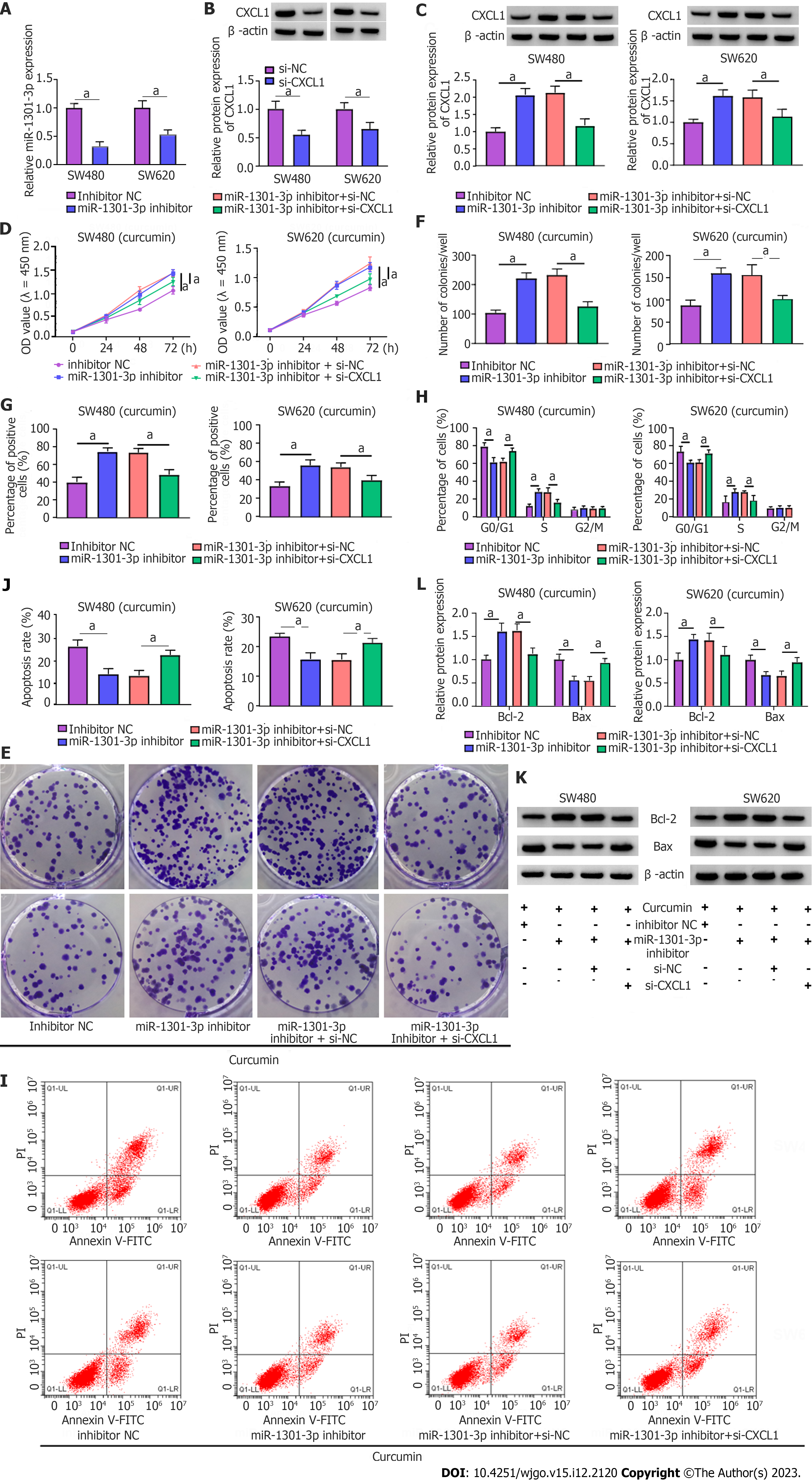
Figure 7 Downregulating CXCL1 abrogated the influence of miR-1301-3p knockdown on curcumin-triggered colorectal carcinoma cell proliferation and apoptosis.
A: Real-time quantitative PCR assay measurement of miR-1301-3p in inhibitor NC and miR-1301-3p inhibitor-transfected SW480 and SW620; B: Western blot assay of CXCL1 protein in si-NC or si-CXCL1-transfected SW480 and SW620; C: Western blot assay of CXCL1 protein in SW480 and SW620 after inhibitor NC, miR-1301-3p inhibitor, miR-1301-3p inhibitor + si-NC and miR-1301-3p inhibitor + si-CXCL1 transfection; D-L: Colorectal carcinoma cells were intervened by curcumin after transfection; D: Cell counting kit-8 assay of cell viability; E and F: Colony-forming cell assay of colony number; G: 5-Ethynyl-2’-deoxyuridine assay of positive cells; H: Flow cytometry assay of cell cycle distribution; I and J: Flow cytometry assay of apoptosis; K and L: Western blot assay of Bcl-2 and Bax protein levels. 1P < 0.05.
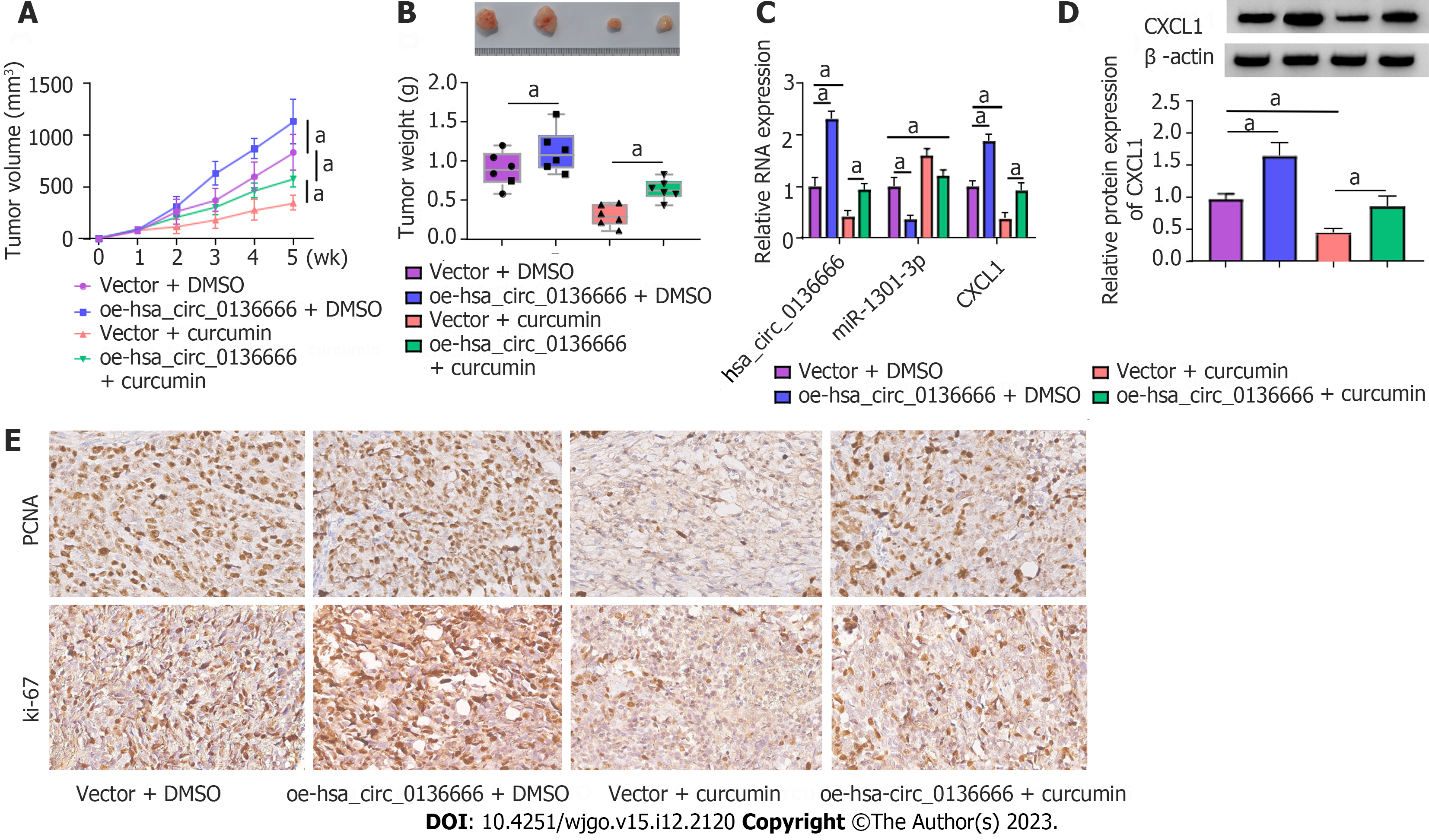
Figure 8 Overexpressing hsa_circ_0136666 abolished the repression of curcumin on colorectal carcinoma growth in vivo.
The nude mice were subcutaneously inoculated with SW480 introduced with vector or oe-hsa_circ_0136666, followed by intraperitoneal curcumin (25 mg/kg) injection one week later. A and B: Tumour volume and tumour weight measurements in the xenografts; C: Real-time quantitative PCR assay quantification of hsa_circ_0136666, miR-1301-3p, and CXCL1 in xenografts; D: Western blot assay of CXCL1 protein in xenografts; E: Immunohistochemical staining of Ki-67 and proliferating cell nuclear antigen expression in xenografts. 1P < 0.05. PCNA: Proliferating cell nuclear antigen.
- Citation: Chen S, Li W, Ning CG, Wang F, Wang LX, Liao C, Sun F. Hsa_circ_0136666 mediates the antitumor effect of curcumin in colorectal carcinoma by regulating CXCL1 via miR-1301-3p. World J Gastrointest Oncol 2023; 15(12): 2120-2137
- URL: https://www.wjgnet.com/1948-5204/full/v15/i12/2120.htm
- DOI: https://dx.doi.org/10.4251/wjgo.v15.i12.2120


