Copyright
©The Author(s) 2022.
World J Gastrointest Oncol. Mar 15, 2022; 14(3): 664-677
Published online Mar 15, 2022. doi: 10.4251/wjgo.v14.i3.664
Published online Mar 15, 2022. doi: 10.4251/wjgo.v14.i3.664
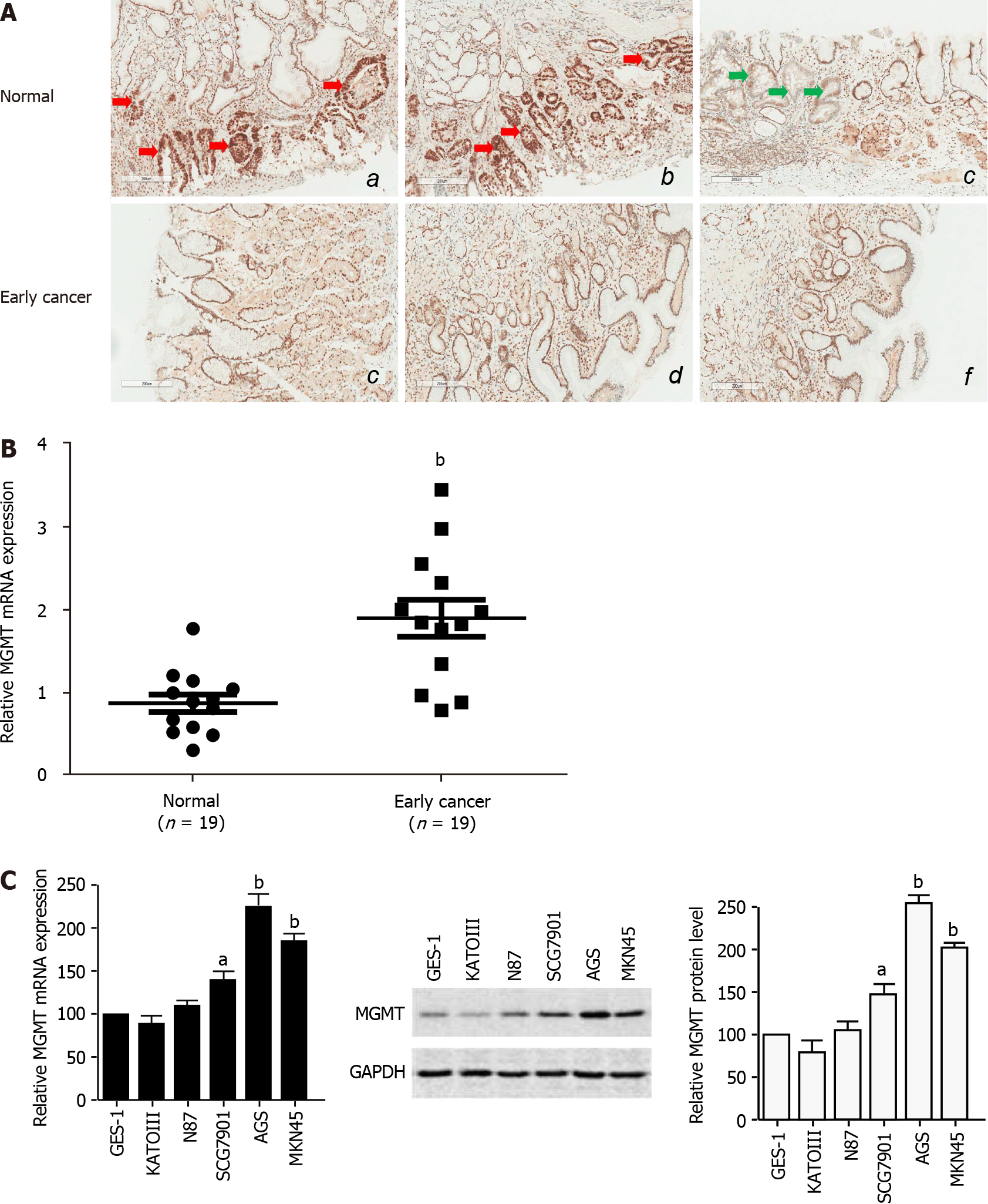
Figure 1 O6-methylguanine-DNA methyltransferase expression is enhanced in early stage gastric cancer.
A: Representative images of immunohistochemistry staining for O6-methylguanine-DNA methyltransferase (MGMT) in early stage gastric tumor and normal tissues (n = 19); B: The mRNA level of MGMT in early stage gastric tumor and adjacent normal tissues (n = 19). The mRNA expression was normalized by glyceraldehyde-3-phosphate dehydrogenase (GAPDH); C: MGMT mRNA and protein expression in normal gastric epithelial cells and cancer cells by quantitative real-time polymerase chain reaction and immunoblot assays. GAPDH was used to normalize MGMT expression. The analyses were repeated three times, and the results are expressed as the mean ± SD. aP < 0.05; bP < 0.01. MG-C: MNNG-induced malignant transformed cell; MU-C: MNU-induced malignant transformed cell; GAPDH: Glyceraldehyde-3-phosphate dehydrogenase.
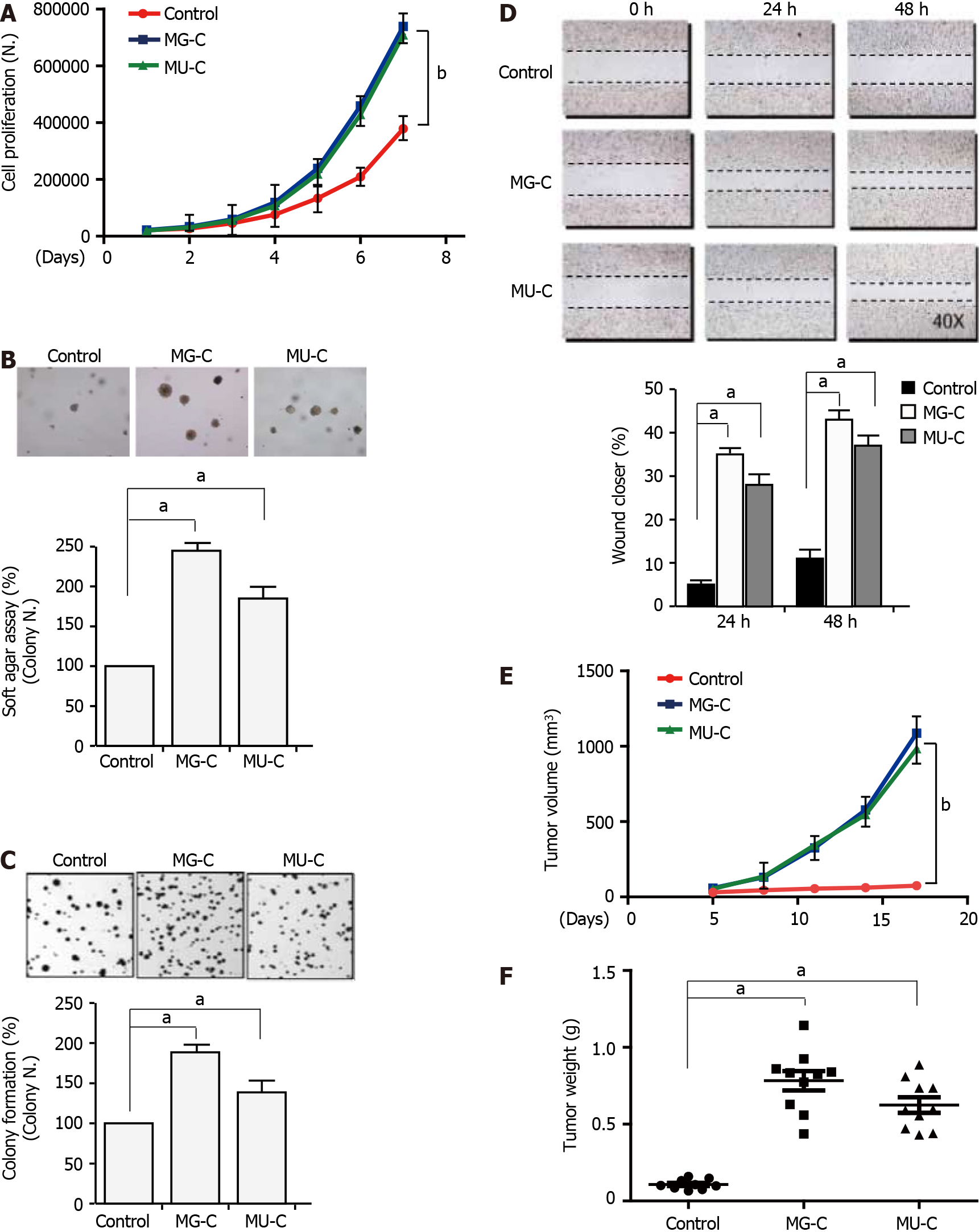
Figure 2 N-nitroso compound treatment induces gastric epithelial cell malignant transformation.
A: Cell proliferation monitored by cell counting in N-methyl-N’-nitro-N-nitrosoguanidine (MNNG)/N-methyl-N-nitroso-urea (MNU)-treated and control cells; B and C: Cell anchorage-independent growth on soft agar and cell colony formation. Top, representative images; bottom, quantitative results of cell colony per field; D: Wound healing assay. Top, representative images of wound healing assay; right, relative percentage of wound closure after treatment; E and F: Tumor growth curve and tumor weight in nude mice injected subcutaneously with the transformed cells induced by MNNG/MNU and control cells. The analyses were repeated three times, and the results are expressed as the mean ± SD. aP < 0.05; bP < 0.01.
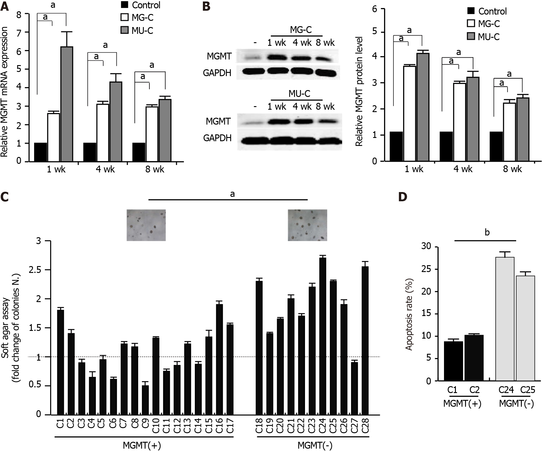
Figure 3 O6-methylguanine-DNA methyltransferase is downregulated in N-nitroso compound-induced gastric epithelial cell malignant transformation.
A and B: O6-methylguanine-DNA methyltransferase (MGMT) mRNA and protein expression in transformed gastric epithelial cells induced by N-methyl-N’-nitro-N-nitrosoguanidine (MNNG)/N-methyl-N-nitroso-urea (MNU) for 1, 4, and 8 wk; C: Cell anchorage-independent growth on soft agar for subcolones of MNNG/MNU-induced cells. C1-28: Different subcolones of MNNG/MNU-induced cells; MGMT(+): MGMT expression is upregulated in these subcolones; MGMT(-): MGMT expression is downregulated or no-changed in these subcolones; D: Apoptosis assay of MNNG/MNU-transformed subcolones after doxycycline treatment. The analyses were repeated three times, and the results are expressed as the mean ± SD. aP < 0.05; bP < 0.01. GAPDH: Glyceraldehyde-3-phosphate dehydrogenase.
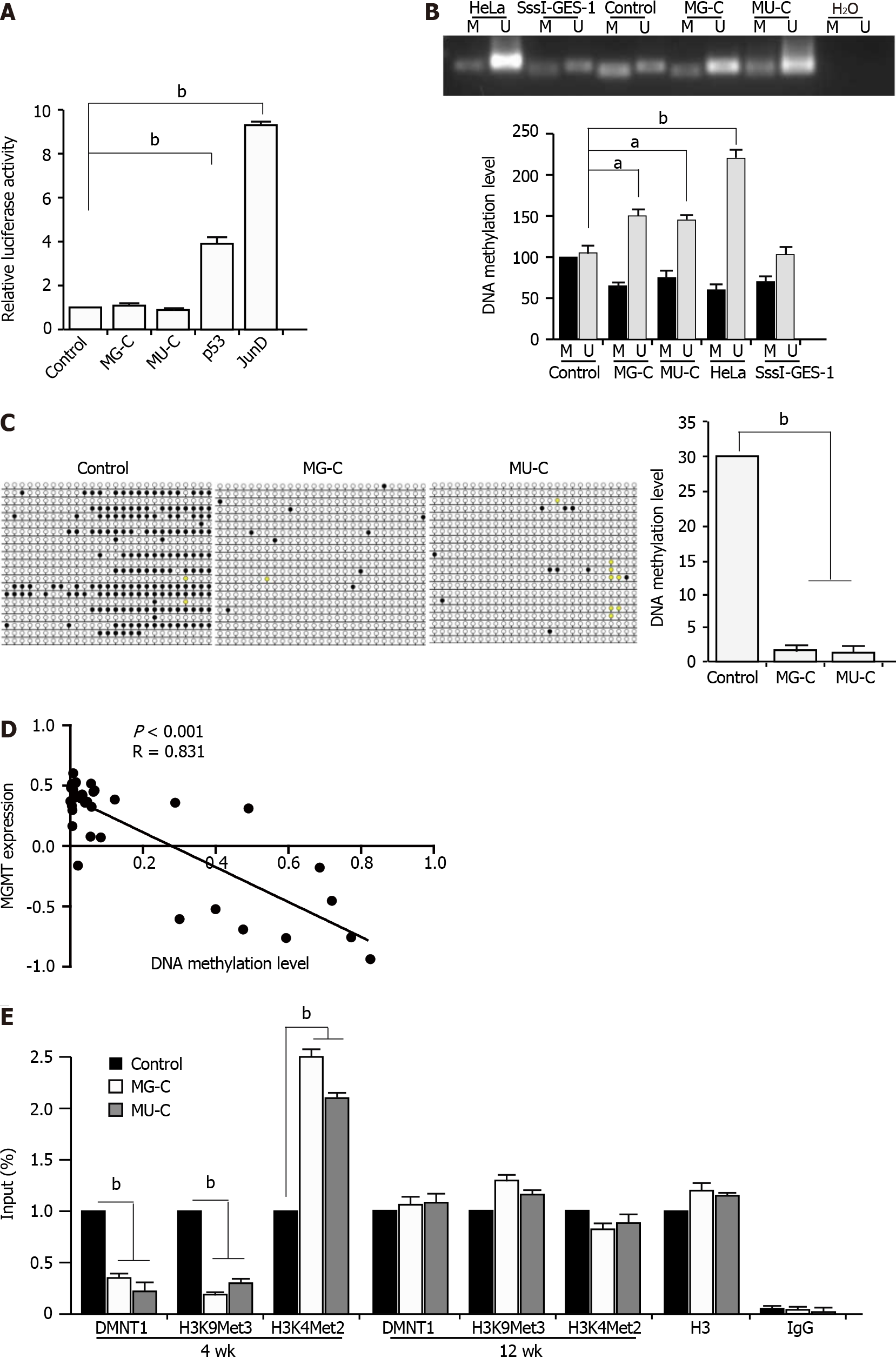
Figure 4 DNA hypomethylation contributes to O6-methylguanine-DNA methyltransferase upregulation in cell malignant transformation.
A: Luciferase reporter assay in control and N-nitroso compound-transformed cells using PGL3-O6-methylguanine-DNA methyltransferase (MGMT) promoter; B and C: Methylation specific polymerase chain reaction and bisulfite genomic sequence analysis of the DNA methylation level of N-methyl-N’-nitro-N-nitrosoguanidine/N-methyl-N-nitroso-urea-induced transformed cells compared with control cells; D: Correlation of MGMT expression and DNA methylation level of MGMT promoter based on the CCLE database; E: ChIP assay with anti-DNMT1 and anti-H3K9Me3 and H3K4Me2 antibodies for analyzing the DNMT1 binding to the MGMT promoter and the H3K9Me3 and H3K4Me2 levels in the MGMT promoter. The analyses were repeated three times, and the results are expressed as the mean ± SD. aP < 0.05; bP < 0.01. M: Methylated; U: Unmethylated; IgG: Immunoglobulin G.
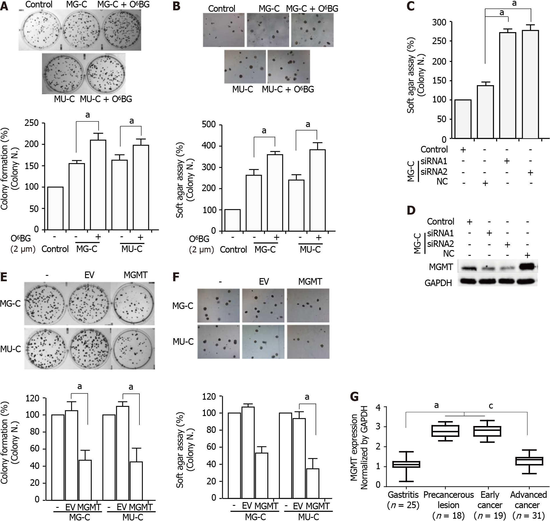
Figure 5 Inhibition of O6-methylguanine-DNA methyltransferase contributes to the N-nitroso compound-induced cell malignant phenotype.
A and B: Cell anchorage-independent growth on soft agar and cell colony formation of N-methyl-N’-nitro-N-nitrosoguanidine/N-methyl-N-nitroso-urea-induced cells after O6-BG treatment; C: Cell anchorage-independent growth on soft agar of cells with O6-methylguanine-DNA methyltransferase (MGMT) knock-down; D: Knock-down efficiency of MGMT detected by Western blot; E and F: Cell anchorage-independent growth on soft agar and cell colony formation of MGMT overexpressing cells; G: The mRNA expression of MGMT in gastric endoscopic biopsy samples. The analyses were repeated three times, and the results are expressed as the mean ± SD. a,cP < 0.05. cP < 0.05, precancerous lesion and early cancer vs advanced cancer. EV: Empty vector; MGMT: MGMT overexpression; MGMT: O6-methylguanine-DNA methyltransferase.
- Citation: Chen YX, He LL, Xiang XP, Shen J, Qi HY. O6-methylguanine DNA methyltransferase is upregulated in malignant transformation of gastric epithelial cells via its gene promoter DNA hypomethylation. World J Gastrointest Oncol 2022; 14(3): 664-677
- URL: https://www.wjgnet.com/1948-5204/full/v14/i3/664.htm
- DOI: https://dx.doi.org/10.4251/wjgo.v14.i3.664


