Copyright
©The Author(s) 2022.
World J Gastrointest Oncol. Dec 15, 2022; 14(12): 2415-2421
Published online Dec 15, 2022. doi: 10.4251/wjgo.v14.i12.2415
Published online Dec 15, 2022. doi: 10.4251/wjgo.v14.i12.2415
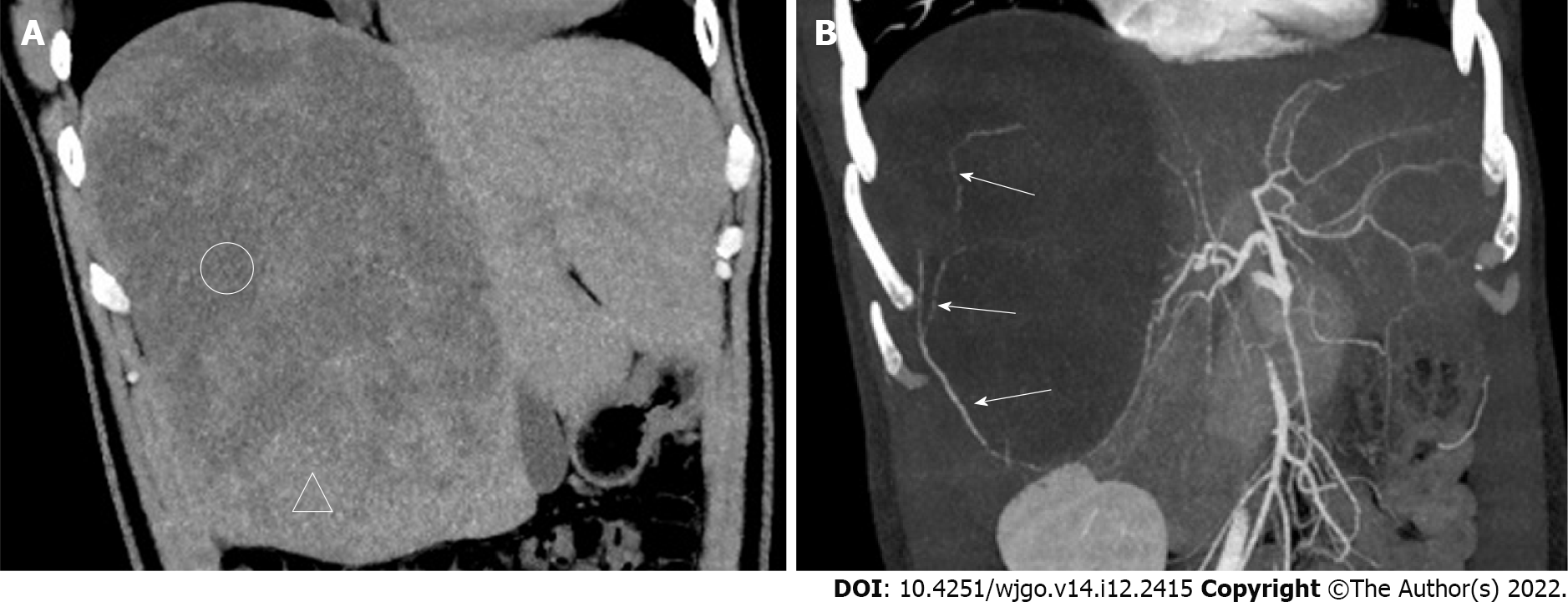
Figure 1 Computed tomography images.
A: Plain computed tomography (CT) coronary reconstruction image; B: CT-enhanced arterial phase maximum intensity projection (MIP) reconstruction image. Compared with the liver parenchyma, many areas of the lesions appear as low density (circled areas) on CT. The mass resembles a meteorite. The MIP image shows that the tumor-feeding artery originates from the right posterior hepatic artery, from bottom to top, with multiple branches (white long arrows in Figure 1B).
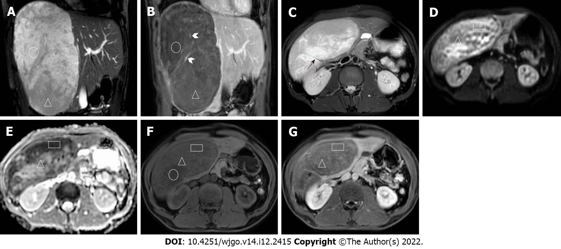
Figure 2 Magnetic resonance imaging images.
A: Coronal balanced fast field echo (B-FFE) image; B: Coronal T1-weighted image (T1WI) after 150-s gadolinium enhancement; C: Axial image, including T2-weighted imaging (T2WI); D: Diffusion-weighted imaging (DWI); E: Apparent diffusion coefficient (ADC); F: T1WI; G: After 7-min gadolinium enhancement, the lesions showed a high signal on B-FFE, a low signal on T1WI, and no definitive enhancement (circled areas). In the lesions, a strip-shaped flow void vascular signal (black short arrow in Figure 2C) can be seen on T2WI. Multiple irregular strip-like obvious enhancement foci with irregular shapes are shown (white arrowhead in Figure 2B). Some areas of high signal on DWI and low signal on ADC and B-FFE (triangular areas) showing progressive and heterogeneous enhancement with gadolinium contrast enhancement. The mass resembled different meteorites in the various sequences.
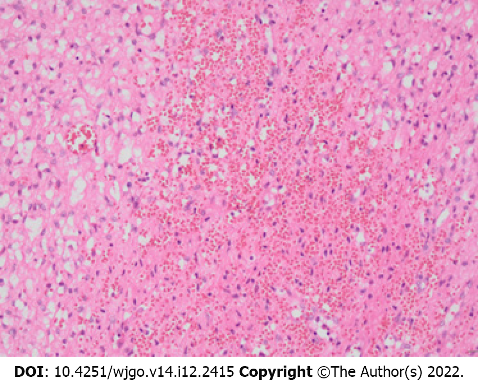
Figure 3 Pathology map (200 ×, hematoxylin-eosin staining).
The tumor was mainly composed of capillaries and eosinophilic, vacuole-containing stromal cells. The cell morphology was mild, the nuclei were small and uniform, and division was rare. The blood vessels were full of blood, with some blood spilling out from the blood vessels.
- Citation: Li DF, Guo XJ, Song SP, Li HB. Rare massive hepatic hemangioblastoma: A case report. World J Gastrointest Oncol 2022; 14(12): 2415-2421
- URL: https://www.wjgnet.com/1948-5204/full/v14/i12/2415.htm
- DOI: https://dx.doi.org/10.4251/wjgo.v14.i12.2415


