Copyright
©The Author(s) 2020.
World J Gastrointest Oncol. Mar 15, 2020; 12(3): 358-364
Published online Mar 15, 2020. doi: 10.4251/wjgo.v12.i3.358
Published online Mar 15, 2020. doi: 10.4251/wjgo.v12.i3.358
Figure 1 Case 1.
A: Positron emission tomography scan: the white arrow shows the area of multiple foci of fludeoxyglucose uptake in the treated area; B: Multi-phase magnetic resonance imaging scan: it shows no arterial enhancement in the same area during the arterial phase; C: Positron emission tomography scan: the arrow indicates an area of fludeoxyglucose uptake, indicating another residual tumor.
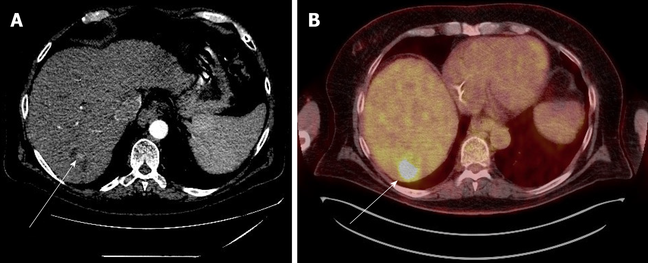
Figure 2 Case 2.
A: Multi-phase computed tomography scan: a hypodense area, indicated by the white arrow, in the liver during the arterial phase post trans-arterial chemo-embolization treatment; B: Positron emission tomography scan: in the same area, there is avid fludeoxyglucose uptake, indicating residual tumor.
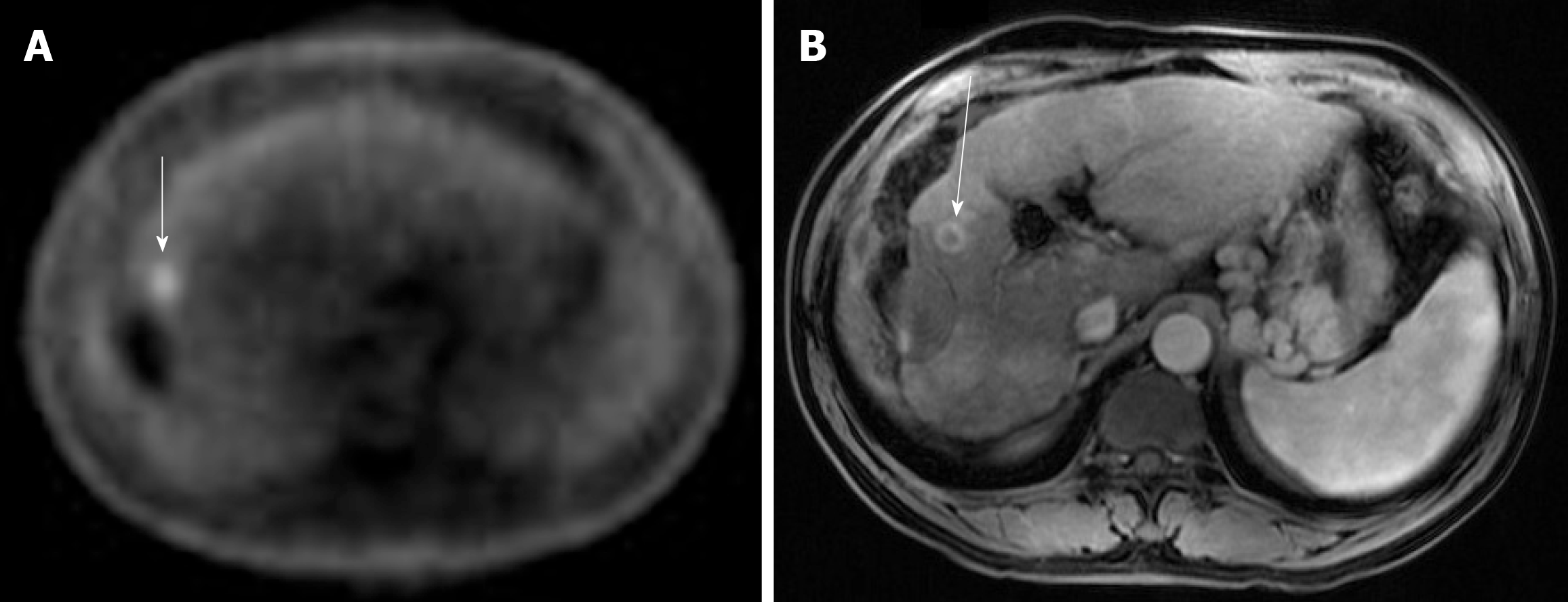
Figure 3 Case 3.
A: Positron emission tomography scan: the white arrow indicates a small focus of fludeoxyglucose uptake adjacent to the treatment zone showing absent metabolic activity; B: Multi-phase magnetic resonance imaging: the white arrow points to focal bleed at the periphery of the treatment zone post microwave ablation without evidence of any viable tumor during the arterial phase.
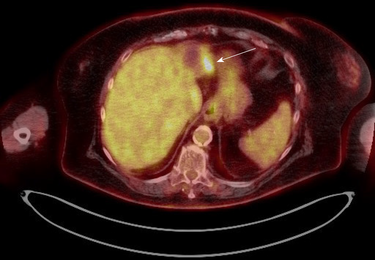
Figure 4 Case 4.
Positron emission tomography scan: the white arrow points to the fludeoxyglucose uptake at the periphery of a treated lesion.
- Citation: Cheng JT, Tan NE, Volk ML. Utility of positron emission tomography-computed tomography scan in detecting residual hepatocellular carcinoma post treatment: Series of case reports. World J Gastrointest Oncol 2020; 12(3): 358-364
- URL: https://www.wjgnet.com/1948-5204/full/v12/i3/358.htm
- DOI: https://dx.doi.org/10.4251/wjgo.v12.i3.358


