Copyright
©The Author(s) 2019.
World J Gastrointest Oncol. Jul 15, 2019; 11(7): 509-517
Published online Jul 15, 2019. doi: 10.4251/wjgo.v11.i7.509
Published online Jul 15, 2019. doi: 10.4251/wjgo.v11.i7.509
Figure 1 Creation of gastric cancer organoids.
Endoscopic biopsy and surgical specimens are obtained from the patient and washed and centrifuged extensively. Endoscopic biopsy specimens are then placed on a microscope slide and pressure is applied by a coverslip onto the tissues to release gastric glands. Surgical specimens are enzymatically digested and minced with a scalpel for gland isolation. Once isolated, glands are suspended in matrigel and plated in drops in pre-warmed wells. Organoids are overlaid with medium.
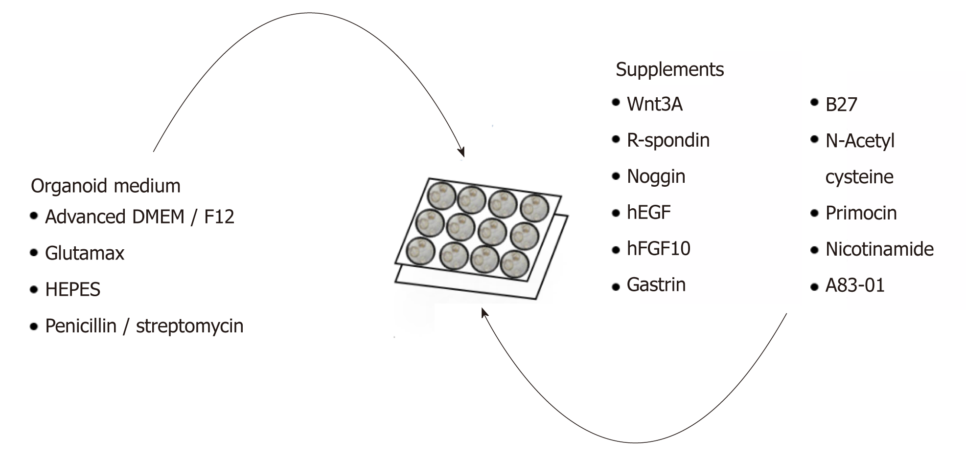
Figure 2 Complete list of ingredients for gastric cancer organoid medium.
Organoid medium consists mainly of advanced DMEM/F12, Glutamax, HEPES, and penicillin/streptomycin and further supplemented with growth factors and enzymes to promote organoid growth.
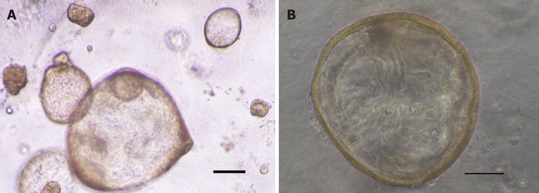
Figure 3 Image of gastric cancer organoids.
A: Image of passage 2 gastric cancer organoids created from biopsy. Scale bar 200 μm; B: High magnification image of passage 2 gastric cancer organoid. Scale bar 100 μm.
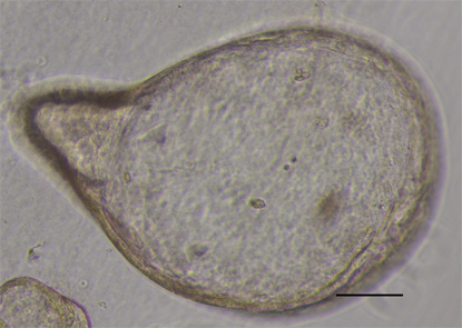
Figure 4 Gastric cancer organoid with budding.
High magnification image of passage 2 gastric cancer organoid with budding. Scale bar 100 μm.
- Citation: Lin M, Gao M, Cavnar MJ, Kim J. Utilizing gastric cancer organoids to assess tumor biology and personalize medicine. World J Gastrointest Oncol 2019; 11(7): 509-517
- URL: https://www.wjgnet.com/1948-5204/full/v11/i7/509.htm
- DOI: https://dx.doi.org/10.4251/wjgo.v11.i7.509


