Published online Jul 10, 2015. doi: 10.4253/wjge.v7.i8.806
Peer-review started: November 2, 2014
First decision: December 12, 2014
Revised: March 26, 2015
Accepted: April 10, 2015
Article in press: April 12, 2015
Published online: July 10, 2015
Processing time: 258 Days and 16.9 Hours
Hilar biliary strictures are caused by various benign and malignant conditions. It is difficult to differentiate benign and malignant strictures. Postcholecystectomy benign biliary strictures are frequently encountered. Endoscopic management of these strictures is challenging. An endoscopic method has been advocated that involves placement of increasing number of stents at regular intervals to resolve the stricture. Malignant hilar strictures are mostly unresectable at the time of diagnosis and only palliation is possible.Endoscopic palliation is preferred over surgery or radiological intervention. Magnetic resonance cholangiopancreaticography is quite important in the management of these strictures. Metal stents are superior to plastic stents. The opinion is divided over the issue of unilateral or bilateral stenting.Minimal contrast or no contrast technique has been advocated during endoscopic retrograde cholangiopancreatography of these patients. The role of intraluminal brachytherapy, intraductal ablation devices, photodynamic therapy, and endoscopic ultrasound still remains to be defined.
Core tip: Management of benign or malignant hilar biliary strictures is difficult. Surgery is technically demanding for benign hilar biliary strictures and results of endoscopic management are not very satisfactory.Endoscopic palliation is preferred modality of managing malignant hilar strictures. However, it is still controversial to drain unilaterally or bilaterally. Use of contrast during endoscopic retrograde cholangiopancreatography and leaving some ducts undrained is a major problem in these patients. We have reviewed the literature on all these aspects of hilar biliary strictures.
- Citation: Singh RR, Singh V. Endoscopic management of hilar biliary strictures. World J Gastrointest Endosc 2015; 7(8): 806-813
- URL: https://www.wjgnet.com/1948-5190/full/v7/i8/806.htm
- DOI: https://dx.doi.org/10.4253/wjge.v7.i8.806
Biliary strictures at hepatic hilum are not uncommon and present a difficult diagnostic and therapeutic problem. Hilar strictures can be benign or malignant which are often difficult to differentiate. Various modalities as surgery, endoscopy and radiology have been used in the management of these strictures with variable results. The role of some newer modalities, e.g., intraluminal brachytherapy, intraductal ablation devices, photodynamic therapy (PDT), still remain investigational.
Etiologically, these strictures can be divided into benign or malignant causes[1] (Table 1). The differentiation of benign from malignant hilar strictures is difficult.
| Malignant hilar strictures |
| Primary tumors (cholangiocarcinoma) |
| Local extension (gallbladder cancer, hepatocellular carcinoma, and pancreatic cancer) |
| Lymph node metastases (Breast, colon, stomach, ovaries, lymphoma, and melanoma) |
| Benign hilar strictures |
| Postoperative injuries (cholecystectomy, liver transplantation, liver resection, and biliodigestive anastomosis) |
| Primary sclerosing cholangitis |
| Others (stone disease, follicular cholangitis, parasite infection, granular cell tumor, chronic fibroinflammatory process, compression from portal cavernomatosis, granulomatous process, and lymphoplasmacyticscleros ingpancreatitis/cholangitis) |
The alkaline phosphatase isoenzyme and CA19-9 have been used to discriminate benign from malignant strictures with variable sensitivity and specificity[2-4]. Radiologic evaluation of patients with hilar strictures can be done with ultrasonography, contrast-enhanced computed tomography (CT) scan, magnetic resonance imaging (MRI) and magnetic resonance cholangiopancreaticography (MRCP). These modalities can help to delineate the level of bilary obstruction along with the extent of biliary dilatation. Any mass lesion or distant metastasis can also be detected with these methods[5-9]. Increased alkaline phosphatase and CA19-9 levels, increased thickness of bile duct wall to ≥ 5 mm and regional lymphadenopathy (> 1 cm) on CT scan, and cholangiographic appearance of abrupt cutoff and separation of biliary ductal system suggest a malignant etiology of hilar obstruction[10]. Another study showed an association of raised bilirubin levels of > 8.4 mg% and CA19-9 level > 100 U/L with malignant etiologies of biliary obstruction[11]. MRI/MRCP performed better than CT to differentiate benign and malignant causes of biliary obstruction[11]. However, in a study of 49 patients with mass lesion at hilum on abdominal ultrasonography or CT scan, raised CEA and CA 19-9 levels, and presence of irregular, eccentric strictures with abrupt cutoff suggesting malignancy on cholangiography, benign diseases was documented in 24% of cases on surgical histopathology[12]. Endoscopic retrograde cholangiopancreatography (ERCP) and percutaneous transhepatic cholangiography (PTC) provide a better assessment of the biliary tree. It also enables brush biopsy and cytology studies providing a histological diagnosis in these patients. However, these procedures carry a significant risk of complications[13,14]. Various studies have shown a variable sensitivity (37%-70%) and high specificity (> 95%) for biopsies or brush cytology (Table 2)[15-18].
Endoscopic ultrasound (EUS) has shown promising results for the diagnosis of hilar strictures. In a study of 24 patients with negative or unsuccessful brush cytology results in cases of proximal bile duct obstruction, EUS revealed a mass lesion in 23 patients (96%)[19]. EUS-FNA in these patients provided a sensitivity of 77% and accuracy of 79%. However, the negative predictive value was quite low (29%). In another study of 44 patients with negative brush cytology in cases of suspected hilarcholangiocarcinoma, EUS-FNA had an accuracy of 91% with 89% sensitivity and 100% specificity[20]. Intraductal ultrasonography enhances the diagnostic accuracy of ERC (88%) as compared to MRC (58%) or ERC alone (76%)[21].
Benign biliary strictures at the hepatic hilum most commonly result from surgical injuries, most often after cholecystectomy. Post-cholecystectomy strictures develop in 0.2%-0.5% of patients undergoing surgery and account for 80% of benign hilar strictures[22]. Post-liver transplant strictures develop in 5.9% of patients[23].
The management and outcome of postsurgical strictures depends on the type of stricture. Bismuth and Lazorthes classified postsurgical biliary strictures based on the level of healthy biliary mucosa suitable for anastomosis (Table 3)[24,25].
| Type I: Common hepatic or main bile duct stump ≥ 2 cm |
| Type II: Common hepatic duct stump < 2 cm |
| Type III: Hilar stricture- ceiling of the biliary confluence is intact, right and left ductal system communicate |
| Type IV: Ceiling of the confluence is destroyed, bile ducts are separated |
| Type V: Type I, II or III plus stricture of an isolated right duct |
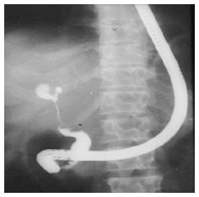
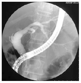
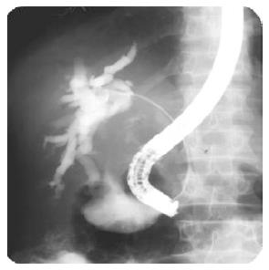
Surgical management of postsurgical benign biliary strictures carries a morbidity of 18%-51%, mortality of 4%-13%, and recurrence rate of 10%-30%[22]. It, therefore, requires specific skills and expertise. The management of hilar postsurgical strictures (types III, IV, and V) is more challenging and results in worse outcomes (Figures 1 and 2)[1]. A retrospective study of 57 patients surgically treated for cicatricial biliary strictures showed a higher rate (14%) of stricture recurrence and cholangitis in patients with hilar obstruction compared to none in patient with lesions below the hilum[26]. A recent study reported the safety and efficacy of right hemihepatectomy with cholangiojejunostomy in patients with strictures involving secondary bile duct branches associated with vascular injuries. The study showed 100% survival without stricture recurrence after a mean follow up of 80 mo. No major postoperative complications were documented[27].
Endoscopic management of postsurgical strictures is more safe and efficacious. Two 10F plastic stents are placed in for a maximum duration of 12 mo. Stent exchange is done at 3 mo interval to reduce the risk of stent blockage and cholangitis (classical approach)[28,29]. Endoscopic management is feasible in 80% of cases. Stricture recurrence occurs in 20% of patients after stent removal over a period of 9.1 years. All the instances of restenosis were noted within 2 years of stent removal. Mean time from stent removal to symptom onset was 2.6 mo (range 1 wk–2 year)[29].
An aggressive approach involves insertion of an increasing number of plastic stents until resolution of stricture, with stent exchange performed at 3-5 mo interval[30]. In a study of 40 patients with 18 hilar strictures, overall success rate with this approach was 89%. Recurrence occurred in only one patient after a mean follow up of 48.8 mo (range 2-11.3 years). Mean number of stents used was 3.2 ± 1.3 (range 1-6) over a period of 12.1 ± 5.3 mo (range 2-24 mo)[30].
Cholangiocarcinoma, carcinoma gall bladder (GB) and secondaries account for majority of malignant hilar biliary strictures. Malignant hilar biliary strictures are classified as per the Bismuth Classification (Table 4)[31]. It carries a poor prognosis with 5 year survival of < 10%. Curative resection is feasible in < 10%. Palliation remains the mainstay of therapy. However, surgical palliation is associated with an unacceptable 33% mortality[31,32].
| Type I: Obstruction within 1 cm of bifurcation but confluence patent |
| Type II: Obstruction limited to confluence |
| Type III: Obstruction at confluence with proximal extension to right or left side |
| Type IV: Obstruction involving bilateral secondary or tertiary branches or multifocal strictures |
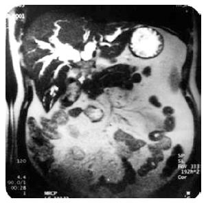
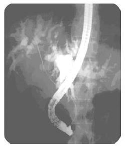
Current options for palliation include surgical bypass, percutaneous drainage and endoscopic stenting.Endoscopic drainage is safer and more successful with a lower propensity to bile leak, infection and haemorrhage. However, a recent randomized controlled study of 54 patients with unresectable carcinoma GB with Bismuth type 2 (Figure 3) or 3 hilar block showed better drainage (89% vs 41%) and lower complication rate (cholangitis 48% vs 11%) with percutaneous approach[33]. Both the groups had similar procedure-related mortality (4% vs 8%), 30-d mortality (4% vs 8%) and median survival (60 d in both; P = 0.71). Percutaneous drainage resulted in a significantly better quality of life, as assessed at 3 mo after the procedure[33]. This study used plastic stents instead of metal stents in unresectable carcinoma GB with Bismuth type 2 or 3 hilar block and biliary ducts were left opacified and undrained after contrast injection which could be responsible for higher rates of complication with endoscopic approach. Hence, the results need to be interpreted with caution.
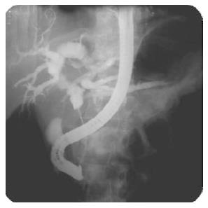
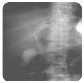
Endoscopic stenting in hilar obstructions can be done with plastic or metal stents (Figures 4-7). Plastic stents are less expensive, have technically easy insertion with relatively easy removal and exchange. But, they have limited stent patency. Metal stents have prolonged stent patency, do not occlude side branches and have easier passage across biliary strictures due to relatively smaller delivery system. But, greater cost and difficulty in removal once blocked are the limitations[34].
Metal stents have been shown to perform better than even large bore plastic endoprostheses. A prospective randomized trial of 20 patients with Bismuth type II-IV hilar obstruction compared 14 French plastic stents with 24 French metal endoprostheses in the management of malignant hilar obstructions with obstructive jaundice[35]. Metal stents insertion was associated with greater success as well as patency rates compared to placement of plastic stent. It was also cost-effective due to lower number of re-interventions required in these patients. Another randomized controlled trial of 108 patients with Bismuth type II-IV unresectable hilarcholangiocarcinoma demonstrated better drainage and more prolonged survival with self-expandable metal stents compared to plastic stents[36]. A meta-analysis of 10 trials showed a significantly higher successful drainage rate [odds ratio (OR) = 0.26; 95%CI: 0.16-0.42; I2 = 40.3%], lower early complication rate (OR = 2.92; 95%CI: 1.65-5.17; I2 = 0%), longer stent patency [hazard ratio (HR) 0.43; 95%CI: 0.30-0.61; I2 = 57.6%], and longer patient survival (HR = 0.73; 95%CI: 0.56-0.96; I2 = 56.9%) with metal stents in comparison with plastic stents[37].
There is much controversy regarding the placement of unilateral or bilateral stents for hilar strictures. In a study of 190 patients with Bismuth type I-III hilar strictures, successful drainage after single stent was achieved in 80% of patients[38]. The placement of a second stent was considered only in patients with new onset cholangitis or incomplete resolution of cholestatic symptoms. Early complications were observed in 7%, 14% and 31% patients with type I, II and III strictures, respectively. De Palma et al[39,40] showed that unilateral stenting is feasible, safe and effective. In a prospective study of 61 patients with hilar malignancy, the placement of a single metal stent across the stricture into duct easier to access achieved successful stent insertion in 96.7% and successful drainage in 96.7% patients. Median survival of these patients was 140 d with median stent patency of 169 d. Stent malfunction was seen in 4.9%[40]. A recent meta-analysis also revealed that unilateral and bilateral biliary drainage may have equivalent efficacy in hilar biliary obstruction with a higher success rate for unilateral stent placement[37]. A case series of 151 patients with unresectable Bismuth type II and III hilar biliary obstruction revealed similar successful drainage rate, complications, 30-d mortality, number of re-interventions and survival based on whether right or left biliary ductal system was drained[41]. However, in patients with bilobar opacification of biliary ductal system, bilateral drainage should be obtained to reduce the risk of cholangitis[42].
It was widely held that draining 25% of liver volume provides adequate palliation of obstructive jaundice with biochemical improvement in these patients[43]. However, a recent study of 107 patients with Bismuth type II-IV hilar strictures concluded that drainage of more than 50% of liver volume predicts efficacy of drainage and translates into longer survival (119 d vs 59 d, P = 0.005),especially in Bismuth type III hilar strictures[44]. Bilateral stent insertion is often required to achieve more than 50% drainage. The study, however, has several drawbacks. The study had a retrospective design, most of the patients underwent plastic instead of metal stenting in hilar biliary obstruction and majority of the patients had cholangiocarcinoma which has relatively prolonged survival and can confound the results.
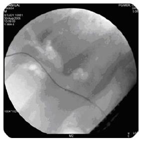
Failure to drain the hepatic lobes or segments after contrast injection is responsible for most of the cases of post procedure early cholangitis and mortality[42]. It brought to the fore the concept of contrast free stenting[45,46]. To avoid bilateral contrast injection and stent placement in Bismuth-type III and IV Klatskin tumors, Hintze et al[47] used MRCP in 35 patients for ERC and unilateral stent insertion. The placement of stents under MRCP guidance reduced incidence of post-ERC bacterial cholangitis. A prospective study of 18 patients with Bismuth type II malignant hilar biliary obstruction demonstrated successful endoscopic drainage in all the patients and no cholangitis or 30-d mortality with MRCP guided contrast-free unilateral metal stenting[45]. Another study of 15 patients with Bismuth type II malignant hilar strictures used contrast-free balloon-assisted unilateral plastic stenting with 100% successful drainage and no cholangitis or 30-d mortality[48]. Comparison of air and iodine contrast cholangiography in hilar strictures in a retrospective study showed less cholangitis with air contrast in Bismuth type II- IV strictures[49]. Subsequently, two studies showed 100% successful stent placement and drainage with no cholangitis and 30 d mortality with contrast-free air cholangiography-assisted (Figure 8) unilateral stent deployment[50,51]. A recent randomized controlled study compared CO2 cholangiography with iodine contrast cholangiography in 36 patients with Bismuth type II-IV malignant hilar obstruction and revealed lower incidence of cholangitis in CO2 group (5.6% vs 33.3%, P = 0.04)[52].
Novel approaches including drug eluting stents, EUS guided biliary drainage, intraluminal brachytherapy, intraductal ablation devices and PDT have recently been used with variable results.
External beam irradiation therapy in malignant biliary strictures is limited by radiation tolerance of liver, bowel and kidneys. Intraluminal brachytherapy allows greater radiation dose locally administered to predefined volume of tissue. It can be administered via endoscopic or percutaneous route. A few recent studies have documented the safety and efficacy of intraluminal brachytherapy in association with stent placement in unresectable, malignant hilar strictures. This new method resulted in prolonged survival in these patients[53-56].
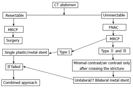
PDT is a promising mode of therapy for unresectable cholangiocarcinoma. It uses a combination of photosensitising chemical and light of appropriate wavelength to generate cytotoxic reactive oxygen species culminating in tumour cell death by necrosis or apoptosis. Continuous biliary drainage is achieved by stent implantation after the procedure. A randomized controlled study of 39 patients with histologically confirmed unresectable cholangiocarcinoma was terminated prematurely due to prolongation of survival (median 493 d vs 98 d; P < 0.0001), more effective biliary drainage and improved quality of life with stenting and PDT in comparison with stenting alone[57]. In a recent retrospective study of 184 patients with hilar cholangiocarcinoma managed with either surgery (60), stenting (56) or stenting with PDT (68); PDT had a longer survival compared to stenting (12.0 mo vs 6.4 mo, P < 0.01) and comparable survival to R1/R2 resection (12.2 mo)[58]. In conclusion,management of hilar biliary strictures is a difficult problem. Surgical, endoscopic and percutaneous approaches have been used in the management of these strictures with variable results. However, the appropriate management also depends on expertise available at that centre. Endoscopic management with increasing number of plastic stents and surgery for failed cases in benign biliary strictures is a reasonable approach. Appropriate management algorithm for malignant hilar strictures is given in Figure 9.
| 1. | Larghi A, Tringali A, Lecca PG, Giordano M, Costamagna G. Management of hilar biliary strictures. Am J Gastroenterol. 2008;103:458-473. [PubMed] |
| 2. | Paritpokee N, Tangkijvanich P, Teerasaksilp S, Wiwanitkit V, Lertmaharit S, Tosukhowong P. Fast liver alkaline phosphatase isoenzyme in diagnosis of malignant biliary obstruction. J Med Assoc Thai. 1999;82:1241-1246. [PubMed] |
| 3. | Akdoğan M, Parlak E, Kayhan B, Balk M, Saydam G, Sahin B. Are serum and biliary carcinoembryonic antigen and carbohydrate antigen19-9 determinations reliable for differentiation between benign and malignant biliary disease. Turk J Gastroenterol. 2003;14:181-184. [PubMed] |
| 4. | Mann DV, Edwards R, Ho S, Lau WY, Glazer G. Elevated tumour marker CA19-9: clinical interpretation and influence of obstructive jaundice. Eur J Surg Oncol. 2000;26:474-479. [RCA] [PubMed] [DOI] [Full Text] [Cited by in Crossref: 186] [Cited by in RCA: 216] [Article Influence: 8.3] [Reference Citation Analysis (0)] |
| 5. | Rösch T, Meining A, Frühmorgen S, Zillinger C, Schusdziarra V, Hellerhoff K, Classen M, Helmberger H. A prospective comparison of the diagnostic accuracy of ERCP, MRCP, CT, and EUS in biliary strictures. Gastrointest Endosc. 2002;55:870-876. [RCA] [PubMed] [DOI] [Full Text] [Cited by in Crossref: 217] [Cited by in RCA: 187] [Article Influence: 7.8] [Reference Citation Analysis (0)] |
| 6. | Kim MJ, Mitchell DG, Ito K, Outwater EK. Biliary dilatation: differentiation of benign from malignant causes--value of adding conventional MR imaging to MR cholangiopancreatography. Radiology. 2000;214:173-181. [RCA] [PubMed] [DOI] [Full Text] [Cited by in Crossref: 113] [Cited by in RCA: 91] [Article Influence: 3.5] [Reference Citation Analysis (0)] |
| 7. | Kehagias D, Metafa A, Hatziioannou A, Mourikis D, Vourtsi A, Prahalias A, Smyrniotis V, Gouliamos A, Vlahos L. Comparison of CT, MRI and CT during arterial portography in the detection of malignant hepatic lesions. Hepatogastroenterology. 2000;47:1399-1403. [PubMed] |
| 8. | Campbell WL, Ferris JV, Holbert BL, Thaete FL, Baron RL. Biliary tract carcinoma complicating primary sclerosing cholangitis: evaluation with CT, cholangiography, US, and MR imaging. Radiology. 1998;207:41-50. [RCA] [PubMed] [DOI] [Full Text] [Cited by in Crossref: 91] [Cited by in RCA: 64] [Article Influence: 2.3] [Reference Citation Analysis (0)] |
| 9. | Lee WJ, Lim HK, Jang KM, Kim SH, Lee SJ, Lim JH, Choo IW. Radiologic spectrum of cholangiocarcinoma: emphasis on unusual manifestations and differential diagnoses. Radiographics. 2001;21 Spec No:S97-S116. [RCA] [PubMed] [DOI] [Full Text] [Cited by in Crossref: 99] [Cited by in RCA: 75] [Article Influence: 3.0] [Reference Citation Analysis (0)] |
| 10. | Kim HJ, Lee KT, Kim SH, Lee JK, Lim JH, Paik SW, Rhee JC. Differential diagnosis of intrahepatic bile duct dilatation without demonstrable mass on ultrasonography or CT: benign versus malignancy. J Gastroenterol Hepatol. 2003;18:1287-1292. [RCA] [PubMed] [DOI] [Full Text] [Cited by in Crossref: 21] [Cited by in RCA: 18] [Article Influence: 0.8] [Reference Citation Analysis (0)] |
| 11. | Saluja SS, Sharma R, Pal S, Sahni P, Chattopadhyay TK. Differentiation between benign and malignant hilar obstructions using laboratory and radiological investigations: a prospective study. HPB (Oxford). 2007;9:373-382. [RCA] [PubMed] [DOI] [Full Text] [Cited by in Crossref: 61] [Cited by in RCA: 69] [Article Influence: 3.6] [Reference Citation Analysis (0)] |
| 12. | Koea J, Holden A, Chau K, McCall J. Differential diagnosis of stenosing lesions at the hepatic hilus. World J Surg. 2004;28:466-470. [RCA] [PubMed] [DOI] [Full Text] [Cited by in Crossref: 43] [Cited by in RCA: 41] [Article Influence: 1.9] [Reference Citation Analysis (0)] |
| 13. | Bilbao MK, Dotter CT, Lee TG, Katon RM. Complications of endoscopic retrograde cholangiopancreatography (ERCP). A study of 10,000 cases. Gastroenterology. 1976;70:314-320. [PubMed] |
| 14. | Sherman S, Ruffolo TA, Hawes RH, Lehman GA. Complications of endoscopic sphincterotomy. A prospective series with emphasis on the increased risk associated with sphincter of Oddi dysfunction and nondilated bile ducts. Gastroenterology. 1991;101:1068-1075. [PubMed] |
| 15. | Venu RP, Geenen JE, Kini M, Hogan WJ, Payne M, Johnson GK, Schmalz MJ. Endoscopic retrograde brush cytology. A new technique. Gastroenterology. 1990;99:1475-1479. [PubMed] |
| 16. | Foutch PG, Kerr DM, Harlan JR, Manne RK, Kummet TD, Sanowski RA. Endoscopic retrograde wire-guided brush cytology for diagnosis of patients with malignant obstruction of the bile duct. Am J Gastroenterol. 1990;85:791-795. [PubMed] |
| 17. | Ferrari Júnior AP, Lichtenstein DR, Slivka A, Chang C, Carr-Locke DL. Brush cytology during ERCP for the diagnosis of biliary and pancreatic malignancies. Gastrointest Endosc. 1994;40:140-145. [RCA] [PubMed] [DOI] [Full Text] [Cited by in Crossref: 146] [Cited by in RCA: 131] [Article Influence: 4.1] [Reference Citation Analysis (0)] |
| 18. | Singh V, Bhasin S, Nain CK, Gupta SK, Singh G, Bose SM. Brush cytology in malignant biliary obstruction. Indian J Pathol Microbiol. 2003;46:197-200. [PubMed] |
| 19. | DeWitt J, Misra VL, Leblanc JK, McHenry L, Sherman S. EUS-guided FNA of proximal biliary strictures after negative ERCP brush cytology results. Gastrointest Endosc. 2006;64:325-333. [RCA] [PubMed] [DOI] [Full Text] [Cited by in Crossref: 162] [Cited by in RCA: 152] [Article Influence: 7.6] [Reference Citation Analysis (0)] |
| 20. | Fritscher-Ravens A, Broering DC, Knoefel WT, Rogiers X, Swain P, Thonke F, Bobrowski C, Topalidis T, Soehendra N. EUS-guided fine-needle aspiration of suspected hilar cholangiocarcinoma in potentially operable patients with negative brush cytology. Am J Gastroenterol. 2004;99:45-51. [RCA] [PubMed] [DOI] [Full Text] [Cited by in Crossref: 202] [Cited by in RCA: 169] [Article Influence: 7.7] [Reference Citation Analysis (0)] |
| 21. | Domagk D, Wessling J, Reimer P, Hertel L, Poremba C, Senninger N, Heinecke A, Domschke W, Menzel J. Endoscopic retrograde cholangiopancreatography, intraductal ultrasonography, and magnetic resonance cholangiopancreatography in bile duct strictures: a prospective comparison of imaging diagnostics with histopathological correlation. Am J Gastroenterol. 2004;99:1684-1689. [PubMed] |
| 22. | Costamagna G, Shah SK, Tringali A. Current management of postoperative complications and benign biliary strictures. Gastrointest Endosc Clin N Am. 2003;13:635-648, ix. [RCA] [PubMed] [DOI] [Full Text] [Cited by in Crossref: 64] [Cited by in RCA: 61] [Article Influence: 2.7] [Reference Citation Analysis (0)] |
| 23. | Thuluvath PJ, Atassi T, Lee J. An endoscopic approach to biliary complications following orthotopic liver transplantation. Liver Int. 2003;23:156-162. [RCA] [PubMed] [DOI] [Full Text] [Cited by in Crossref: 135] [Cited by in RCA: 133] [Article Influence: 5.8] [Reference Citation Analysis (0)] |
| 25. | Bismuth H, Majno PE. Biliary strictures: classification based on the principles of surgical treatment. World J Surg. 2001;25:1241-1244. [RCA] [PubMed] [DOI] [Full Text] [Cited by in Crossref: 186] [Cited by in RCA: 174] [Article Influence: 7.0] [Reference Citation Analysis (0)] |
| 26. | Monteiro da Cunha JE, Machado MC, Herman P, Bacchella T, Abdo EE, Penteado S, Jukemura J, Montagnini A, Machado MA, Pinotti HW. Surgical treatment of cicatricial biliary strictures. Hepatogastroenterology. 1998;45:1452-1456. [PubMed] |
| 27. | Sugawara G, Ebata T, Yokoyama Y, Igami T, Mizuno T, Nagino M. Management strategy for biliary stricture following laparoscopic cholecystectomy. J Hepatobiliary Pancreat Sci. 2014;21:889-895. [RCA] [PubMed] [DOI] [Full Text] [Cited by in Crossref: 5] [Cited by in RCA: 7] [Article Influence: 0.6] [Reference Citation Analysis (0)] |
| 28. | Davids PH, Rauws EA, Coene PP, Tytgat GN, Huibregtse K. Endoscopic stenting for post-operative biliary strictures. Gastrointest Endosc. 1992;38:12-18. [RCA] [PubMed] [DOI] [Full Text] [Cited by in Crossref: 92] [Cited by in RCA: 81] [Article Influence: 2.4] [Reference Citation Analysis (0)] |
| 29. | Bergman JJ, Burgemeister L, Bruno MJ, Rauws EA, Gouma DJ, Tytgat GN, Huibregtse K. Long-term follow-up after biliary stent placement for postoperative bile duct stenosis. Gastrointest Endosc. 2001;54:154-161. [RCA] [PubMed] [DOI] [Full Text] [Cited by in Crossref: 124] [Cited by in RCA: 101] [Article Influence: 4.0] [Reference Citation Analysis (0)] |
| 30. | Costamagna G, Pandolfi M, Mutignani M, Spada C, Perri V. Long-term results of endoscopic management of postoperative bile duct strictures with increasing numbers of stents. Gastrointest Endosc. 2001;54:162-168. [RCA] [PubMed] [DOI] [Full Text] [Cited by in Crossref: 299] [Cited by in RCA: 272] [Article Influence: 10.9] [Reference Citation Analysis (0)] |
| 31. | Bismuth H, Castaing D, Traynor O. Resection or palliation: priority of surgery in the treatment of hilar cancer. World J Surg. 1988;12:39-47. [RCA] [PubMed] [DOI] [Full Text] [Cited by in Crossref: 233] [Cited by in RCA: 239] [Article Influence: 6.3] [Reference Citation Analysis (0)] |
| 32. | Blumgart LH, Hadjis NS, Benjamin IS. Surgery and hepatic duct carcinoma. Lancet. 1984;1:795. [RCA] [PubMed] [DOI] [Full Text] [Cited by in RCA: 1] [Reference Citation Analysis (0)] |
| 33. | Saluja SS, Gulati M, Garg PK, Pal H, Pal S, Sahni P, Chattopadhyay TK. Endoscopic or percutaneous biliary drainage for gallbladder cancer: a randomized trial and quality of life assessment. Clin Gastroenterol Hepatol. 2008;6:944-950.e3. [RCA] [PubMed] [DOI] [Full Text] [Cited by in Crossref: 98] [Cited by in RCA: 102] [Article Influence: 5.7] [Reference Citation Analysis (0)] |
| 34. | Lee TH. Technical tips and issues of biliary stenting, focusing on malignant hilar obstruction. Clin Endosc. 2013;46:260-266. [RCA] [PubMed] [DOI] [Full Text] [Full Text (PDF)] [Cited by in Crossref: 21] [Cited by in RCA: 25] [Article Influence: 1.9] [Reference Citation Analysis (0)] |
| 35. | Wagner HJ, Knyrim K, Vakil N, Klose KJ. Plastic endoprostheses versus metal stents in the palliative treatment of malignant hilar biliary obstruction. A prospective and randomized trial. Endoscopy. 1993;25:213-218. [RCA] [PubMed] [DOI] [Full Text] [Cited by in Crossref: 234] [Cited by in RCA: 235] [Article Influence: 7.1] [Reference Citation Analysis (0)] |
| 36. | Sangchan A, Kongkasame W, Pugkhem A, Jenwitheesuk K, Mairiang P. Efficacy of metal and plastic stents in unresectable complex hilar cholangiocarcinoma: a randomized controlled trial. Gastrointest Endosc. 2012;76:93-99. [RCA] [PubMed] [DOI] [Full Text] [Cited by in Crossref: 150] [Cited by in RCA: 188] [Article Influence: 13.4] [Reference Citation Analysis (0)] |
| 37. | Hong W, Sun X, Zhu Q. Endoscopic stenting for malignant hilar biliary obstruction: should it be metal or plastic and unilateral or bilateral. Eur J Gastroenterol Hepatol. 2013;25:1105-1112. [RCA] [PubMed] [DOI] [Full Text] [Cited by in Crossref: 29] [Cited by in RCA: 40] [Article Influence: 3.1] [Reference Citation Analysis (0)] |
| 38. | Polydorou AA, Cairns SR, Dowsett JF, Hatfield AR, Salmon PR, Cotton PB, Russell RC. Palliation of proximal malignant biliary obstruction by endoscopic endoprosthesis insertion. Gut. 1991;32:685-689. [RCA] [PubMed] [DOI] [Full Text] [Cited by in Crossref: 181] [Cited by in RCA: 172] [Article Influence: 4.9] [Reference Citation Analysis (0)] |
| 39. | De Palma GD, Galloro G, Siciliano S, Iovino P, Catanzano C. Unilateral versus bilateral endoscopic hepatic duct drainage in patients with malignant hilar biliary obstruction: results of a prospective, randomized, and controlled study. Gastrointest Endosc. 2001;53:547-553. [RCA] [PubMed] [DOI] [Full Text] [Cited by in Crossref: 336] [Cited by in RCA: 295] [Article Influence: 11.8] [Reference Citation Analysis (1)] |
| 40. | De Palma GD, Pezzullo A, Rega M, Persico M, Patrone F, Mastantuono L, Persico G. Unilateral placement of metallic stents for malignant hilar obstruction: a prospective study. Gastrointest Endosc. 2003;58:50-53. [RCA] [PubMed] [DOI] [Full Text] [Cited by in Crossref: 143] [Cited by in RCA: 130] [Article Influence: 5.7] [Reference Citation Analysis (0)] |
| 41. | Polydorou AA, Chisholm EM, Romanos AA, Dowsett JF, Cotton PB, Hatfield AR, Russell RC. A comparison of right versus left hepatic duct endoprosthesis insertion in malignant hilar biliary obstruction. Endoscopy. 1989;21:266-271. [RCA] [PubMed] [DOI] [Full Text] [Cited by in Crossref: 78] [Cited by in RCA: 72] [Article Influence: 1.9] [Reference Citation Analysis (0)] |
| 42. | Chang WH, Kortan P, Haber GB. Outcome in patients with bifurcation tumors who undergo unilateral versus bilateral hepatic duct drainage. Gastrointest Endosc. 1998;47:354-362. [RCA] [PubMed] [DOI] [Full Text] [Cited by in Crossref: 311] [Cited by in RCA: 272] [Article Influence: 9.7] [Reference Citation Analysis (1)] |
| 43. | Dowsett JF, Vaira D, Hatfield AR, Cairns SR, Polydorou A, Frost R, Croker J, Cotton PB, Russell RC, Mason RR. Endoscopic biliary therapy using the combined percutaneous and endoscopic technique. Gastroenterology. 1989;96:1180-1186. [PubMed] |
| 44. | Vienne A, Hobeika E, Gouya H, Lapidus N, Fritsch J, Choury AD, Chryssostalis A, Gaudric M, Pelletier G, Buffet C. Prediction of drainage effectiveness during endoscopic stenting of malignant hilar strictures: the role of liver volume assessment. Gastrointest Endosc. 2010;72:728-735. [RCA] [PubMed] [DOI] [Full Text] [Cited by in Crossref: 186] [Cited by in RCA: 228] [Article Influence: 14.3] [Reference Citation Analysis (0)] |
| 45. | Singh V, Singh G, Verma GR, Singh K, Gulati M. Contrast-free unilateral endoscopic palliation in malignant hilar biliary obstruction: new method. J Gastroenterol Hepatol. 2004;19:589-592. [RCA] [PubMed] [DOI] [Full Text] [Cited by in Crossref: 18] [Cited by in RCA: 17] [Article Influence: 0.8] [Reference Citation Analysis (0)] |
| 46. | De Palma GD, Lombardi G, Rega M, Simeoli I, Masone S, Siciliano S, Maione F, Salvatori F, Balzano A, Persico G. Contrast-free endoscopic stent insertion in malignant biliary obstruction. World J Gastroenterol. 2007;13:3973-3976. [RCA] [PubMed] [DOI] [Full Text] [Full Text (PDF)] [Cited by in CrossRef: 4] [Cited by in RCA: 4] [Article Influence: 0.2] [Reference Citation Analysis (0)] |
| 47. | Hintze RE, Abou-Rebyeh H, Adler A, Veltzke-Schlieker W, Felix R, Wiedenmann B. Magnetic resonance cholangiopancreatography-guided unilateral endoscopic stent placement for Klatskin tumors. Gastrointest Endosc. 2001;53:40-46. [RCA] [PubMed] [DOI] [Full Text] [Cited by in Crossref: 129] [Cited by in RCA: 114] [Article Influence: 4.6] [Reference Citation Analysis (0)] |
| 48. | Singh V, Singh G, Verma GR, Gupta V, Gupta R, Kapoor R. Contrast-free Balloon-assisted Unilateral Plastic Stenting in Malignant Hilar Biliary Obstruction: A new method. Digestive Endoscopy. 2008;20:190-193. [RCA] [DOI] [Full Text] [Cited by in Crossref: 1] [Cited by in RCA: 2] [Article Influence: 0.1] [Reference Citation Analysis (0)] |
| 49. | Pisello F, Geraci G, Modica G, Sciumè C. Cholangitis prevention in endoscopic Klatskin tumor palliation: air cholangiography technique. Langenbecks Arch Surg. 2009;394:1109-1114. [RCA] [PubMed] [DOI] [Full Text] [Cited by in Crossref: 18] [Cited by in RCA: 15] [Article Influence: 0.9] [Reference Citation Analysis (0)] |
| 50. | Singh V, Singh G, Gupta V, Gupta R, Kapoor R. Contrast-free air cholangiography-assisted unilateral plastic stenting in malignant hilar biliary obstruction. Hepatobiliary Pancreat Dis Int. 2010;9:88-92. [PubMed] |
| 51. | Sud R, Puri R, Hussain S, Kumar M, Thawrani A. Air cholangiogram: a new technique for biliary imaging during ERCP. Gastrointest Endosc. 2010;72:204-208. [RCA] [PubMed] [DOI] [Full Text] [Cited by in Crossref: 12] [Cited by in RCA: 14] [Article Influence: 0.9] [Reference Citation Analysis (0)] |
| 52. | Zhang R, Zhao L, Liu Z, Wang B, Hui N, Wang X, Huang R, Luo H, Fan D, Pan Y. Effect of CO2 cholangiography on post-ERCP cholangitis in patients with unresectable malignant hilar obstruction - a prospective, randomized controlled study. Scand J Gastroenterol. 2013;48:758-763. [RCA] [PubMed] [DOI] [Full Text] [Cited by in Crossref: 6] [Cited by in RCA: 10] [Article Influence: 0.8] [Reference Citation Analysis (0)] |
| 53. | Bruha R, Petrtyl J, Kubecova M, Marecek Z, Dufek V, Urbanek P, Kodadova J, Chodounsky Z. Intraluminal brachytherapy and selfexpandable stents in nonresectable biliary malignancies--the question of long-term palliation. Hepatogastroenterology. 2001;48:631-637. [PubMed] |
| 54. | Veeze-Kuijpers B, Meerwaldt JH, Lameris JS, van Blankenstein M, van Putten WL, Terpstra OT. The role of radiotherapy in the treatment of bile duct carcinoma. Int J Radiat Oncol Biol Phys. 1990;18:63-67. [RCA] [PubMed] [DOI] [Full Text] [Cited by in Crossref: 65] [Cited by in RCA: 61] [Article Influence: 1.7] [Reference Citation Analysis (0)] |
| 55. | Singh V, Kapoor R, Solanki KK, Singh G, Verma GR, Sharma SC. Endoscopic intraluminal brachytherapy and metal stent in malignant hilar biliary obstruction: a pilot study. Liver Int. 2007;27:347-352. [RCA] [PubMed] [DOI] [Full Text] [Cited by in Crossref: 22] [Cited by in RCA: 17] [Article Influence: 0.9] [Reference Citation Analysis (0)] |
| 56. | Aggarwal R, Patel FD, Kapoor R, Kang M, Kumar P, Chander Sharma S. Evaluation of high-dose-rate intraluminal brachytherapy by percutaneous transhepatic biliary drainage in the palliative management of malignant biliary obstruction--a pilot study. Brachytherapy. 2013;12:162-170. [RCA] [PubMed] [DOI] [Full Text] [Cited by in Crossref: 13] [Cited by in RCA: 16] [Article Influence: 1.1] [Reference Citation Analysis (0)] |
| 57. | Ortner ME, Caca K, Berr F, Liebetruth J, Mansmann U, Huster D, Voderholzer W, Schachschal G, Mössner J, Lochs H. Successful photodynamic therapy for nonresectable cholangiocarcinoma: a randomized prospective study. Gastroenterology. 2003;125:1355-1363. [RCA] [PubMed] [DOI] [Full Text] [Cited by in Crossref: 446] [Cited by in RCA: 386] [Article Influence: 16.8] [Reference Citation Analysis (0)] |
| 58. | Witzigmann H, Berr F, Ringel U, Caca K, Uhlmann D, Schoppmeyer K, Tannapfel A, Wittekind C, Mossner J, Hauss J. Surgical and palliative management and outcome in 184 patients with hilar cholangiocarcinoma: palliative photodynamic therapy plus stenting is comparable to r1/r2 resection. Ann Surg. 2006;244:230-239. [RCA] [PubMed] [DOI] [Full Text] [Cited by in Crossref: 235] [Cited by in RCA: 200] [Article Influence: 10.0] [Reference Citation Analysis (0)] |
Open-Access: This article is an open-access article which was selected by an in-house editor and fully peer-reviewed by external reviewers. It is distributed in accordance with the Creative Commons Attribution Non Commercial (CC BY-NC 4.0) license, which permits others to distribute, remix, adapt, build upon this work non-commercially, and license their derivative works on different terms, provided the original work is properly cited and the use is non-commercial. See: http://creativecommons.org/licenses/by-nc/4.0/
P- Reviewer: Tsuyuguchi T S- Editor: Ji FF L- Editor: A E- Editor: Wu HL


