Published online May 16, 2015. doi: 10.4253/wjge.v7.i5.540
Peer-review started: December 16, 2014
First decision: January 20, 2015
Revised: February 9, 2015
Accepted: April 1, 2015
Article in press: April 1, 2015
Published online: May 16, 2015
Processing time: 186 Days and 5.6 Hours
AIM: To compare the characteristics of two single-incision methods, and conventional laparoscopy in cholecystectomy, and demonstrate the safety and feasibility.
METHODS: Three hundred patients with gallstones or gallbladder polyps were admitted to two clinical centers from January 2013 to January 2014 and were randomized into three groups of 100: single-incision three-device group, X-Cone group, and conventional group. The operative time, intraoperative blood loss, complications, postoperative pain, cosmetic score, length of hospitalization, and hospital costs were compared, with a follow-up duration of 1 mo.
RESULTS: A total of 142 males (47%) and 158 females (53%) were enrolled in this study. The population characteristics of these three groups is no significant differences exist in terms of age, sex, body mass index and American Society of Anesthesiology (P > 0.05). In results, there were no significant differences in blood loss, length of hospitalization, postoperative complications.The operative time in X-Cone group was significantly longer than other groups.There were significant differences in postoperative pain scores and cosmetic scores at diffent times after surgery (P < 0.05).
CONCLUSION: This study shows that this two single-incision methods are safe and feasible. Both methods are superior to the conventional procedure in cosmetic and pain scores.
Core tip: This is an article about single-incision laparoscopic surgery. It compares three methods in laparoscopic cholecystectomy. The study concludes that the three-device and X-Cone methods are safe and feasible for single-incision laparoscopic cholecystectomy. Compared with conventional laparoscopic cholecystectomy, single-incision laparoscopic surgery techniques have advantages in pain and cosmetic factors.
- Citation: He GL, Jiang ZS, Cheng Y, Lai QB, Zhou CJ, Liu HY, Gao Y, Pan MX, Jian ZX. Tripartite comparison of single-incision and conventional laparoscopy in cholecystectomy: A multicenter trial. World J Gastrointest Endosc 2015; 7(5): 540-546
- URL: https://www.wjgnet.com/1948-5190/full/v7/i5/540.htm
- DOI: https://dx.doi.org/10.4253/wjge.v7.i5.540
Single-incision laparoscopic surgery (SILS) is an area of research interest in minimally invasive surgery. Its main advantage is a scar-free abdominal wall after surgery, as well as milder postoperative pain, faster recovery, shorter hospital stay, and better cosmetic outcomes. Since the first report of single-incision resection of gallbladder through the abdominal cavity by Navarra et al[1] in 1997, there has been a growing number of clinical reports on this topic[2-10]. At present, a variety of auxiliary means are used, such as the X-Cone method, triport method, Kirschner-aid exposure method, suspension sutures method, and three-device method[11-16]. However, there has been no comparative study of the various methods.
We enrolled 200 cases of laparoscopic cholecystectomy completed using the three-device and X-Cone methods in our two centers, as well as 100 cases of conventional laparoscopic cholecystectomy, to compare their technical characteristics and clinical outcomes, and demonstrate the safety and feasibility of the single-incision methods.
Inclusion criteria were: patients with gallstones or gallbladder polyps; age 18-85 years; either sex; and body mass index (BMI) < 35 kg/m2. Exclusion criteria were: complication by common bile duct or intrahepatic bile duct stones; acute cholecystitis; suspicion of complicated cholecystitis; BMI ≥ 35 kg/m2; drug addiction; ASA physical classification > 3; previous upper abdominal surgery; pregnancy; presence of umbilical hernia; or previous umbilical hernia repair.
All 300 patients were admitted to the two clinical centers for laparoscopic cholecystectomy from January 2013 to January 2014. They were randomly assigned to three groups of 100. The case characteristics are shown in Table 1. All surgery was performed by three surgeons, each of whom had conducted > 1000 cholecystectomies, including ≥ 100 single-incision laparoscopic cholecystectomies.
| X-Cone method(n = 100)(No.1 group) | Three-device method(n = 100)(No.2 group) | Conventional method(n = 100)(No.3 group) | P value | Statistical methods and values | |
| Sex | |||||
| Male | 47 | 44 | 52 | χ2 = 1.31 | |
| Female | 53 | 56 | 48 | ||
| Age (yr) | 39.5 ± 14.5 | 40.0 ± 12.5 | 41.7 ± 12.0 | 0.465 | One-Way ANOVA F = 0.768 |
| BMI (kg/m2) | 26.1 ± 5.5 | 28.2 ± 7.5 | 26.1 ± 8.4 | 0.06 | One-Way ANOVA F = 2.847 |
| Surgical risk grade (ASA) | 1.6 ± 0.5 | 1.6 ± 0.4 | 1.6 ± 0.4 | 0.681 | One-Way ANOVA F = 0.385 |
| Diagnosis | |||||
| Stones | 58 | 52 | 47 | χ2 = 2.43 | |
| Polyps | 42 | 48 | 53 | ||
The primary end points of this study were feasibility and safety of the three-device method and X-Cone method compared with conventional laparoscopic cholecystectomy, as indicated by intraoperative and postoperative adverse events up to 1 mo, operative time, and estimated blood loss. The secondary end points were: (1) pain as determined by a 10-point pain intensity scale performed at days 1 and 2, 1 wk, and 1 mo; (2) cosmesis evaluated via a body image questionnaire, photo series questionnaire, and cosmesis scale performed at 1 and 2 wk, and 1 mo; and (3) length of hospital stay and hospital costs.
Umbilical disinfection was completed 1 d before surgery. Following routine anesthesia with tracheal intubation, second-generation cephalosporin was intraoperatively administered once. After pneumoperitoneum was established in patients undergoing three-device or conventional surgery, the patients were placed with their legs closed in the Trendelenburg position at approximately 30°, left tilted at approximately 20°. The surgeons stood on the left side of the patient, with the monitor on the right side. For patients undergoing X-Cone surgery, the legs were placed apart in the Trendelenburg position at approximately 30°, left tilted approximately 20°. The surgeons stood between the legs with the monitor on the patient’s head side.
General anesthesia was induced with propofol (2 mg/kg) and sufentanil (0.5-2 μg/kg). Tracheal intubation facilitated by injection of Atracurium (0.5 mg/kg). Anesthesia during surgery was maintained with isoflurane 1.2% and administration of Atracurium (0.1 mg/kg) and sufentanil (0.1 μg/kg) and every 30 min. The patients were monitored by ECG, pulse oximetry, noninvasive blood pressure. Patients were recovered by administration of neostigmine (40 μg/kg) and atropine (20 μg/kg).
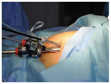
Three-device method: The umbilical incision was approximately 2.0 cm. Three trocars were directly placed into the incision. The locations are shown in Figure 1. The inferior 10-mm trocar was for insertion of the 30° laparoscope, while the two 5-mm trocars above were working ports for the scalpel and forceps, respectively. There was 1-2 mm of tissue between the three trocars to prevent leakage. The cystic artery was directly cut with the ultrasonic scalpel, and the cystic duct was closed with a 5-mm Hem-o-lok titanium clamp and transected with scissors. If the 5-mm Hem-o-lok was too small for the occlusion, the 5-mm trocar in the right working port was replaced with a 10-mm one for placement of a 10-mm Hem-o-lok. Once there was no abnormality of the abdomen, the gallbladder was removed. All equipment was removed first, and a pair of vessel forceps was inserted into the original 10-mm trocar to enlarge the incision in the abdominal cavity, and grasping forceps and a 10-mm trocar laparoscope were in turn placed to extract the gallbladder as a whole. The umbilicus white line was closed with a 3-0 Polysorb absorbable suture, and the umbilical skin incisions intradermally closed with absorbable sutures.
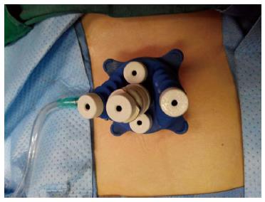
X-Cone method: A 3.0-cm curved incision was made around the upper or lower edge of the umbilicus. The subcutaneous tissue and anterior sheath were divided and the posterior sheath separated. As the middle space was pulled with hemostatic forceps, the X-Cone device (Karl Storz, Tuttlingen, Germany) was inserted (Figure 2). Pneumoperitoneum up to 12 mmHg was established through the pole of the X-Cone, and a 5-mm 30° laparoscope was inserted. The clamp and scalpel were placed into the other two ports. The surgeon pulled the gallbladder with curved traction forceps in the left hand and resected the gallbladder triangle with the ultrasonic scalpel in the right hand. The cystic artery was directly separated with the scalpel. After separation of the cystic duct, a 5 or 10-mm Hem-o-lok was used to close it and the cystic duct was then cut with scissors. The gallbladder was then removed as a whole from the gallbladder bed. The gallbladder was taken directly from the umbilical port. The umbilicus white line was closed with a 3-0 Polysorb absorbable suture, and the umbilical skin incisions intradermally closed with absorbable sutures.
Conventional method: A curved incision of 1.0 cm was made at the umbilical lower edge, an incision of 1.0-1.2 cm was made below the xiphoid, and a 0.5-cm incision was made 1-2 cm above the right clavicular line at the umbilical level. Two 10-mm trocars and one 5-mm trocar were placed into these incisions. The 10-mm umbilical trocar was for placement of the laparoscope, and the other two were working ports for placement of the ultrasonic scalpel and forceps.
Postoperative care: After completion of surgery in all three groups, the incisions were treated with a 50% dose of 75 mg ropivacaine for local anesthesia. Subsequently, the patients were extubated and closely observed in the postanesthetic care unit and then transferred to the surgical ward once their Aldrete score was ≥ 9. Postoperative electrocardiography was performed and oxygen was administered for 6 h, in combination with rehydration and bleeding control, as well as other fluid replacement. Liquid food and ambulation were allowed 6 h after surgery. In the postoperative period, Intravenous rotundine sulfate, at a dose of 1 mg/kg was administered according to patient request every 12 h until discharge home. Surgical dressings were changed on the first day after surgery. The patients were discharged on the second day after surgery. They were also asked to return for check-up at 1, 2 wk and 1 mo after surgery.
Data were analyzed using SPSS version 13 (Chicago, IL, United States). Base on Kolmogorov-Smirnov test, operative time, estimated blood loss, postoperative hospital stay, pain scores and cosmetic scores were all summarized using mean ± SD and compared among the 3 groups by using the One-Way ANOVA test (Tukey method). Intraoperative and postoperative adverse events was compared among the three procedures by Fisher exact test. χ2 tests were performed to explore the effects of sex, and the clinical diagnosis. A value of P < 0.05 was considered to indicate significance.
A total of 300 patients were enrolled in this study and assigned to three groups of 100: three-device, X-Cone method, and conventional method. There were no significant differences in age, sex, BMI and ASA among the groups. The operation time, blood loss and complications are listed in Table 2. There were no significant differences in blood loss and postoperative hospital stay. The X-Cone method required longer operation time compared to the conventional (56.3 min vs 42.1 min, P = 0.000) and three-device methods (56.3 min vs 45.6 min, P = 0.000), while the latter two did not differ significantly in this regard (42.1 min vs 45.6 min, P = 0.111). Hospitalization costs were higher in the X-Cone group than the three-device group (P = 0.000) and the conventional group (P = 0.000). The conventional group was the cheapest group in the three groups.
| X-Cone method(n = 100)(No.1 group) | Three-device method(n = 100)(No.2 group) | Conventional method(n = 100)(No.3 group) | P values | Statistical methodsand values | |
| Operative time (min) | 56.3 ± 14.0 | 45.6 ± 12.0 | 42.1 ± 11.0 | 0.000 G1 vs G2 0.000 G1 vs G3 0.000 G2 vs G3 0.111 | One-Way |
| ANOVA F = 36.86 | |||||
| Blood loss1 (mL) | 16.4 ± 3.7 | 17.1 ± 4.5 | 15.8 ± 4.7 | 0.089 | One-Way ANOVA F = 2.439 |
| Conversion to multiple-incision LC | 1 | 2 | 0 | 0.776 | Fisher exact test |
| Complications | |||||
| Incision contusion | 3 | 4 | 1 | 0.543 | Fisher exact test |
| Wound infection | 1 | 1 | 3 | 0.625 | Fisher exact test |
| Bile duct injury | 0 | 0 | 0 | 1.0 | Fisher exact test |
| Bile leakage | 2 | 2 | 1 | 1.0 | Fisher exact test |
| Abdominal infection | 0 | 0 | 0 | 1.0 | Fisher exact test |
| Postoperative hospital stay (d) | 1.66 ± 0.5 | 1.69 ± 0.5 | 1.68 ± 0.4 | 0.928 | One-Way ANOVA F = 0.075 |
| Hospital costs | 11658 ± 1435 | 10406 ± 1246 | 10036 ± 1154 | 0.000 G1 vs G2 0.000 G1 vs G3 0.000 G2 vs G3 0.415 | One-Way ANOVA F = 52.66 |
In the X-Cone group, there were three cases of surgical incision contusion, and one case of wound hematoma. In the three-device group, two patients required additional working ports due to severe inflammatory adhesions, and there were four cases of incision contusion. In the conventional method group, all patients were successfully operated, and there were one case of incision contusion and three cases of incision wound infection under the xiphoid. No patient converted to laparotomy, and there was no serious complication such as bile duct injury or bile peritonitis. There was no postoperative bleeding or conversion to laparotomy. Percutaneous incision suture was successful without umbilical hernia.
The pain and cosmetic scores are listed in Table 3. The pain score was evaluated using a visual analog scale of 1-10 on days 1, 2 and 7, as well as 1 mo after surgery. There were differences in the pain scores on day 1 between the single-incision methods and the conventional method in favor of the former (P < 0.0001), there was no difference between the two single-incision methods (P = 0.296). The X-Cone group was the most comfortable on day 2, while the three-device group on day 7 after surgery. At 1 mo, single-incision methods were better than the conventional method.
| X-Conemethod(n = 100) (No.1 group) | Three-device method(n = 100)(No.2 group) | Conventional method(n = 100)(No.3 group) | P values | Statistical methodsand values | |
| Pain score1 | One-Way ANOVA | ||||
| 1 d after surgery | 3.4 ± 1.2 | 3.6 ± 1.2 | 4.2 ± 1.1 | 0 G1 vs G2 0.296 G1 vs G3 0.000 G2 vs G3 0.005 | F = 11.16 |
| 2 d after surgery | 2.8 ± 0.8 | 3.0 ± 1.0 | 3.2 ± 1.0 | 0.002 G1 vs G2 0.155 G1 vs G3 0.001 G2 vs G3 0.204 | F = 6.34 |
| 7 d after surgery | 2.2 ± 0.6 | 2.0 ± 0.6 | 2.3 ± 0.7 | 0.014 G1 vs G2 0.252 G1 vs G3 0.365 G2 vs G3 0.010 | F = 4.35 |
| 1 mo after surgery | 1.6 ± 0.4 | 1.5 ± 0.3 | 1.7 ± 0.5 | 0 G1 vs G2 0.123 G1 vs G3 0.048 G2 vs G3 0.000 | F = 9.435 |
| Cosmetic score2 | |||||
| 1 wk after surgery | 8 ± 0.7 | 8 ± 0.5 | 6 ± 0.4 | 0 G1 vs G2 0.999 G1 vs G3 0.000 G2 vs G3 0.000 | F = 423.61 |
| 2 wk after surgery | 8 ± 0.8 | 8 ± 0.6 | 7 ± 0.3 | 0 G1 vs G2 0.966 G1 vs G3 0.000 G2 vs G3 0.000 | F = 93.67 |
| 1 mo after surgery | 9 ± 0.2 | 9 ± 0.3 | 8 ± 0.5 | 0 G1 vs G2 0.814 G1 vs G3 0.000 G2 vs G3 0.000 | F = 308.9 |
The cosmetic scores were rated on a 1–10 scale with questionnaires, with 10 being satisfied and 0 being unsatisfied. At 1 wk (P = 0.000), 2 wk (P = 0.000) and 1 mo (P = 0.000) after surgery, the single-incision methods were significantly better than the conventional group in terms of cosmetic scores. The X-Cone group and the three-device group had no differences (P > 0.05).
SILS techniques have been extensively applied both at home and abroad in recent years[7,11,17-20]. It is performed using a 1-wound laparoscopic surgical procedure or by using speciic ports[21-24].Compared with conventional laparoscopic cholecystectomy, they are associated with fewer injuries and better cosmetic outcomes, as well as many other advantages[25-29]. Some investigators believe that single-incision laparoscopic cholecystectomy will replace conventional laparoscopic cholecystectomy, and become the new gold standard[13,14].
This was an unplanned preliminary analysis of a continuing clinical trial to establish the safety of SILS as an operative approach for treatment of gallbladder disease. This article presents preliminary data of a multicenter, prospective randomized, single-blinded study comparing two single-incision cholecystectomy (three-device and X-Cone methods) with conventional standard multiport laparoscopic cholecystectomy. Primary end points included feasibility and safety, with pain, cosmesis, and costs as secondary end points.
In terms of feasibility and safety, except for the two patients who had additional working ports due to severe inflammatory adhesions in the three-device group, all patients underwent surgery successfully. None of the 200 patients converted to laparotomy or had complications such as bile duct injury, suggesting that single-incision laparoscopic cholecystectomy was feasible and safe. The low conversion rate may differ from that in other studies[18,30], which was probably due to the fact that patients with acute cholecystitis were excluded from our study. There were no significant differences in the complication rates among the three groups. There were four cases of incision contusion in the three-device group, and three and one cases in the X-Cone and conventional groups, respectively. To avoid conflict of instruments in the abdominal cavity with the single-incision method, repeated external squeezing of the surrounding tissue is often required, which may explain the incision contusion in the three-device and X-Cone groups. In addition, there were different numbers of cases of bile leakage in all groups, which were treated with repeated rinsing with saline until the liquid turned clear. There was no case of biliary peritonitis infection afterwards.
There was no significant difference in blood loss and postoperative hospital stay. The X-Cone method required a longer operation time compared to the conventional (56.3 min vs 42.1 min, P = 0.000) and three-device methods (56.3 min vs 45.6 min, P = 0.000). Although all three surgeons had conducted > 100 cases of gallbladder SILS, the X-Cone procedure was associated with inconvenient operation across multiple ports and conflicting handling of instruments such as curved apparatus and solid textures, which might have extended the operation time. In contrast, the three-device method and conventional technique did not differ significantly in this regard (45.6 min vs 42.1 min, P = 0.111). The space between the instruments in the three-device method comprises soft subcutaneous tissue, which allows for a wider range of motion for the instruments, which is conducive to surgery.
Regarding the pain and cosmetic scores, there were differences between the single-incision methods and the conventional method in the pain score on day 1 after surgery, in favor of the single-incision methods. The main complaint was pain below the xiphoid incision in the conventional group. As the pain scores declined on days 2 and 7, as well as 1 mo after surgery, the differences became insignificant. At 1 and 2 wk and 1 mo after surgery, the single-incision methods were significantly better than the conventional group in terms of cosmetic scores. No difference was noted between the three device and X-Cone methods.
There was no difference in the hospitalization costs between the three-device and conventional methods, but there was when compared with the X-Cone method, suggesting that the latter method had an impact on the overall hospital costs. In three-device techniques, conventional equipment and devices were used, resulting in no cost difference from the conventional method, so the three-device method has a more cost-effective. Hence, the three-device approach is more suitable for community hospitals in China.
The present study had the following limitations. First, patients with acute cholecystitis were excluded, and this explains the low laparotomy conversion and low complication rates. Second, although all three surgeons had conducted > 100 operations for gallbladder SILS, the X-Cone procedure was associated with inconvenient operation across multiple ports and conflicting handling of instruments such as curved apparatus and solid textures, which might have extended the operation time. Both of these limitations are routinely seen when a new technique is evaluated. Also, long-term complications were not addressed by this study. The frequency of events still needs to be evaluated by long-term trials.
In summary, both the three-device and X-Cone methods are safe and feasible for single-incision laparoscopic cholecystectomy. Compared with conventional laparoscopic cholecystectomy, SILS techniques have advantages in pain and cosmetic factors. Due to its use of conventional instruments and cost-effective nature, the three-device method is more suitable for community hospitals in China, while the X-Cone device, which allows the placement of more surgical instruments, is more advantageous in more complicated procedures such as laparoscopic liver resection.
Single-incision laparoscopic cholecystectomy is a new laparoscopic procedure in laparoscopic surgery. This technique has been denominated by some authors as “scarless”. The best advantage is a scar-free abdominal wall after surgery, as well as milder postoperative pain, faster recovery, shorter hospital stay, and better cosmetic outcomes.
It is a lot of studies about the single-incision laparoscopic surgery (SILS). But there has been no previous reported study of the comparison of these three methods in cholecystectomy.
In this study, the three-device and X-Cone methods are safe and feasible for single-incision laparoscopic cholecystectomy. Compared with conventional laparoscopic cholecystectomy, SILS techniques have advantages in pain and cosmetic factors.
These two SILS techniques were used more and more in different hospitals. Further study is needed to confirm whether these potential advantages of the SILS techniques can change the clinical course of patients with liver surgery.
This is a very interesting paper about the SILS in cholecystectomy. The most important innovations of this study that was applied in the manuscript is the comparison of three methods in cholecystectomy. In addition, it demonstated that single-incision three-device and X-Cone methods are safe and feasible for laparoscopic cholecystectomy.
P- Reviewers: Amornyotin S, Aytac E, Han HS S- Editor: Ma YJ L- Editor: A E- Editor: Wu HL
| 1. | Navarra G, Pozza E, Occhionorelli S, Carcoforo P, Donini I. One-wound laparoscopic cholecystectomy. Br J Surg. 1997;84:695. [PubMed] |
| 2. | Duron VP, Nicastri GR, Gill PS. Novel technique for a single-incision laparoscopic surgery (SILS) approach to cholecystectomy: single-institution case series. Surg Endosc. 2011;25:1666-1671. [RCA] [PubMed] [DOI] [Full Text] [Cited by in Crossref: 25] [Cited by in RCA: 27] [Article Influence: 1.7] [Reference Citation Analysis (0)] |
| 3. | Chang SK, Tay CW, Bicol RA, Lee YY, Madhavan K. A case-control study of single-incision versus standard laparoscopic cholecystectomy. World J Surg. 2011;35:289-293. [RCA] [PubMed] [DOI] [Full Text] [Cited by in Crossref: 56] [Cited by in RCA: 60] [Article Influence: 4.0] [Reference Citation Analysis (0)] |
| 4. | Koo EJ, Youn SH, Baek YH, Roh YH, Choi HJ, Kim YH, Jung GJ. Review of 100 cases of single port laparoscopic cholecystectomy. J Korean Surg Soc. 2012;82:179-184. [RCA] [PubMed] [DOI] [Full Text] [Full Text (PDF)] [Cited by in Crossref: 11] [Cited by in RCA: 13] [Article Influence: 0.9] [Reference Citation Analysis (0)] |
| 5. | Kroh M, El-Hayek K, Rosenblatt S, Chand B, Escobar P, Kaouk J, Chalikonda S. First human surgery with a novel single-port robotic system: cholecystectomy using the da Vinci Single-Site platform. Surg Endosc. 2011;25:3566-3573. [RCA] [PubMed] [DOI] [Full Text] [Cited by in Crossref: 153] [Cited by in RCA: 127] [Article Influence: 8.5] [Reference Citation Analysis (0)] |
| 6. | Roberts KE, Solomon D, Duffy AJ, Bell RL. Single-incision laparoscopic cholecystectomy: a surgeon’s initial experience with 56 consecutive cases and a review of the literature. J Gastrointest Surg. 2010;14:506-510. [RCA] [PubMed] [DOI] [Full Text] [Cited by in Crossref: 90] [Cited by in RCA: 95] [Article Influence: 5.9] [Reference Citation Analysis (0)] |
| 7. | Liang P, Huang XB, Zuo GH. Transumbilical single-port laparoscopic cholecystectomy. Shijie Huaren Xiaohua Zazhi. 2010;9:290-291. |
| 8. | Erbella J, Bunch GM. Single-incision laparoscopic cholecystectomy: the first 100 outpatients. Surg Endosc. 2010;24:1958-1961. [RCA] [PubMed] [DOI] [Full Text] [Cited by in Crossref: 105] [Cited by in RCA: 112] [Article Influence: 7.0] [Reference Citation Analysis (0)] |
| 9. | Barband A, Fakhree MB, Kakaei F, Daryani A. Single-incision laparoscopic cholecystectomy using glove port in comparison with standard laparoscopic cholecystectomy SILC using glove port. Surg Laparosc Endosc Percutan Tech. 2012;22:17-20. [RCA] [PubMed] [DOI] [Full Text] [Cited by in Crossref: 13] [Cited by in RCA: 15] [Article Influence: 1.1] [Reference Citation Analysis (0)] |
| 10. | Rivas H, Varela E, Scott D. Single-incision laparoscopic cholecystectomy: initial evaluation of a large series of patients. Surg Endosc. 2010;24:1403-1412. [RCA] [PubMed] [DOI] [Full Text] [Full Text (PDF)] [Cited by in Crossref: 153] [Cited by in RCA: 149] [Article Influence: 9.3] [Reference Citation Analysis (0)] |
| 11. | Pan MX, Cheng Y, Gao Y. “Three-point” transumbilical single-port laparoscopic cholecystectomy. J South Med Univ. 2010;30:1715-1717. |
| 12. | Sharma A, Soni V, Baijal M, Khullar R, Najma K, Chowbey PK. Single port versus multiple port laparoscopic cholecystectomy-a comparative study. Indian J Surg. 2013;75:115-122. [RCA] [PubMed] [DOI] [Full Text] [Cited by in Crossref: 8] [Cited by in RCA: 13] [Article Influence: 0.9] [Reference Citation Analysis (0)] |
| 13. | Emami CN, Garrett D, Anselmo D, Torres M, Nguyen NX. Single-incision laparoscopic cholecystectomy in children: a feasible alternative to the standard laparoscopic approach. J Pediatr Surg. 2011;46:1909-1912. [RCA] [PubMed] [DOI] [Full Text] [Cited by in Crossref: 18] [Cited by in RCA: 22] [Article Influence: 1.5] [Reference Citation Analysis (0)] |
| 14. | Jacob DA, Raakow R. Single-port transumbilical endoscopic cholecystectomy: a new standard? Dtsch Med Wochenschr. 2010;135:1363-1367. [RCA] [PubMed] [DOI] [Full Text] [Cited by in Crossref: 3] [Cited by in RCA: 7] [Article Influence: 0.4] [Reference Citation Analysis (0)] |
| 15. | Piskun G, Rajpal S. Transumbilical laparoscopic cholecystectomy utilizes no incisions outside the umbilicus. J Laparoendosc Adv Surg Tech A. 1999;9:361-364. [RCA] [PubMed] [DOI] [Full Text] [Cited by in Crossref: 325] [Cited by in RCA: 304] [Article Influence: 11.3] [Reference Citation Analysis (0)] |
| 16. | Bessa SS, Al-Fayoumi TA, Katri KM, Awad AT. Clipless laparoscopic cholecystectomy by ultrasonic dissection. J Laparoendosc Adv Surg Tech A. 2008;18:593-598. [RCA] [PubMed] [DOI] [Full Text] [Cited by in Crossref: 39] [Cited by in RCA: 55] [Article Influence: 3.1] [Reference Citation Analysis (0)] |
| 17. | Wong JS, Cheung YS, Fong KW, Chong CC, Lee KF, Wong J, Lai PB. Comparison of postoperative pain between single-incision laparoscopic cholecystectomy and conventional laparoscopic cholecystectomy: prospective case-control study. Surg Laparosc Endosc Percutan Tech. 2012;22:25-28. [RCA] [PubMed] [DOI] [Full Text] [Cited by in Crossref: 16] [Cited by in RCA: 21] [Article Influence: 1.5] [Reference Citation Analysis (0)] |
| 18. | Vidal O, Valentini M, Ginestà C, Espert JJ, Martinez A, Benarroch G, Anglada MT, García-Valdecasas JC. Single-incision versus standard laparoscopic cholecystectomy: comparison of surgical outcomes from a single institution. J Laparoendosc Adv Surg Tech A. 2011;21:683-686. [RCA] [PubMed] [DOI] [Full Text] [Cited by in Crossref: 16] [Cited by in RCA: 18] [Article Influence: 1.2] [Reference Citation Analysis (0)] |
| 19. | Ponsky TA. Single port laparoscopic cholecystectomy in adults and children: tools and techniques. J Am Coll Surg. 2009;209:e1-e6. [RCA] [PubMed] [DOI] [Full Text] [Cited by in Crossref: 43] [Cited by in RCA: 39] [Article Influence: 2.3] [Reference Citation Analysis (0)] |
| 20. | Guo W, Zhang ZT, Han W, Li JS, Jin L, Liu J, Zhao XM, Wang Y. Transumbilical single-port laparoscopic cholecystectomy: a case report. Chin Med J (Engl). 2008;121:2463-2464. [PubMed] |
| 21. | Yeo D, Mackay S, Martin D. Single-incision laparoscopic cholecystectomy with routine intraoperative cholangiography and common bile duct exploration via the umbilical port. Surg Endosc. 2012;26:1122-1127. [RCA] [PubMed] [DOI] [Full Text] [Cited by in Crossref: 24] [Cited by in RCA: 23] [Article Influence: 1.5] [Reference Citation Analysis (0)] |
| 22. | Joseph M, Phillips M, Rupp CC. Single-incision laparoscopic cholecystectomy: a combined analysis of resident and attending learning curves at a single institution. Am Surg. 2012;78:119-124. [PubMed] |
| 23. | Sasaki K, Watanabe G, Matsuda M, Hashimoto M. Single-incision laparoscopic cholecystectomy: comparison analysis of feasibility and safety. Surg Laparosc Endosc Percutan Tech. 2012;22:108-113. [RCA] [PubMed] [DOI] [Full Text] [Cited by in Crossref: 6] [Cited by in RCA: 7] [Article Influence: 0.5] [Reference Citation Analysis (0)] |
| 24. | Arroyo JP, Martín-Del-Campo LA, Torres-Villalobos G. Single-incision laparoscopic cholecystectomy: is it a plausible alternative to the traditional four-port laparoscopic approach? Minim Invasive Surg. 2012;2012:347607. [RCA] [PubMed] [DOI] [Full Text] [Full Text (PDF)] [Cited by in Crossref: 6] [Cited by in RCA: 9] [Article Influence: 0.6] [Reference Citation Analysis (0)] |
| 25. | Leung D, Yetasook AK, Carbray J, Butt Z, Hoeger Y, Denham W, Barrera E, Ujiki MB. Single-incision surgery has higher cost with equivalent pain and quality-of-life scores compared with multiple-incision laparoscopic cholecystectomy: a prospective randomized blinded comparison. J Am Coll Surg. 2012;215:702-708. [RCA] [PubMed] [DOI] [Full Text] [Cited by in Crossref: 418] [Cited by in RCA: 241] [Article Influence: 16.1] [Reference Citation Analysis (0)] |
| 26. | Lee PC, Lo C, Lai PS, Chang JJ, Huang SJ, Lin MT, Lee PH. Randomized clinical trial of single-incision laparoscopic cholecystectomy versus minilaparoscopic cholecystectomy. Br J Surg. 2010;97:1007-1012. [RCA] [PubMed] [DOI] [Full Text] [Cited by in Crossref: 127] [Cited by in RCA: 137] [Article Influence: 8.6] [Reference Citation Analysis (1)] |
| 27. | Lai EC, Yang GP, Tang CN, Yih PC, Chan OC, Li MK. Prospective randomized comparative study of single incision laparoscopic cholecystectomy versus conventional four-port laparoscopic cholecystectomy. Am J Surg. 2011;202:254-258. [PubMed] |
| 28. | Gangl O, Hofer W, Tomaselli F, Sautner T, Függer R. Single incision laparoscopic cholecystectomy (SILC) versus laparoscopic cholecystectomy (LC)-a matched pair analysis. Langenbecks Arch Surg. 2011;396:819-824. [RCA] [PubMed] [DOI] [Full Text] [Cited by in Crossref: 47] [Cited by in RCA: 56] [Article Influence: 3.7] [Reference Citation Analysis (0)] |
| 29. | Pan MX, Jiang ZS, Cheng Y, Xu XP, Zhang Z, Qin JS, He GL, Xu TC, Zhou CJ, Liu HY. Single-incision vs three-port laparoscopic cholecystectomy: prospective randomized study. World J Gastroenterol. 2013;19:394-398. [RCA] [PubMed] [DOI] [Full Text] [Full Text (PDF)] [Cited by in CrossRef: 38] [Cited by in RCA: 51] [Article Influence: 3.9] [Reference Citation Analysis (0)] |
| 30. | Chiruvella A, Sarmiento JM, Sweeney JF, Lin E, Davis SS. Iatrogenic combined bile duct and right hepatic artery injury during single incision laparoscopic cholecystectomy. JSLS. 2010;14:268-271. [RCA] [PubMed] [DOI] [Full Text] [Full Text (PDF)] [Cited by in Crossref: 11] [Cited by in RCA: 13] [Article Influence: 0.8] [Reference Citation Analysis (0)] |


