©2013 Baishideng Publishing Group Co.
World J Gastrointest Endosc. May 16, 2013; 5(5): 261-264
Published online May 16, 2013. doi: 10.4253/wjge.v5.i5.261
Published online May 16, 2013. doi: 10.4253/wjge.v5.i5.261
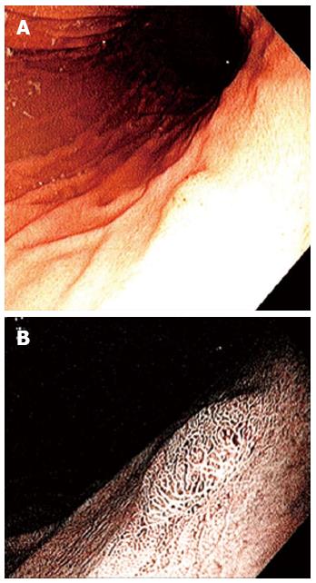
Figure 1 Endoscopic view.
A: Gastric polyp; B: Gastric polyp after indigocarmine staining.
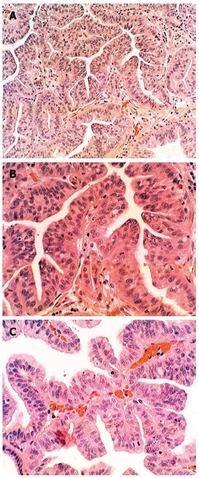
Figure 2 The histological examination of the gastric polypoid lesion revealed a serrated adenoma showing protruding glands with lateral saw tooth-like notches due to scalloped epithelial indentations with high-grade dysplasia.
A: Adenoma showing serrated glands lined with high-grade dysplasia [hematoxylin and eosin (HE) × 10]; B: High power view of the adenomatous component showing serrated glands with indentations lined with high-grade dysplasia (HE × 20); C: View of a single elongated gland with saw-tooth-like configuration lined with high-grade dysplasia (HE × 20).
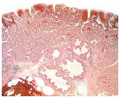
Figure 3 Low-power view of serrated adenoma with invasive carcinoma (hematoxylin and eosin × 10).
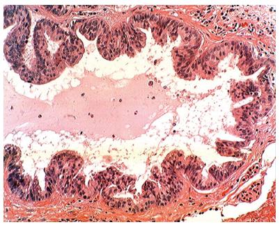
Figure 4 High power view of the invasive component with retained serrated configuration (hematoxylin and eosin × 10).
- Citation: Rubio CA, Björk J. Serrated adenoma of the stomach: Case report and literature review. World J Gastrointest Endosc 2013; 5(5): 261-264
- URL: https://www.wjgnet.com/1948-5190/full/v5/i5/261.htm
- DOI: https://dx.doi.org/10.4253/wjge.v5.i5.261


