Copyright
©2013 Baishideng Publishing Group Co.
World J Gastrointest Endosc. Oct 16, 2013; 5(10): 514-518
Published online Oct 16, 2013. doi: 10.4253/wjge.v5.i10.514
Published online Oct 16, 2013. doi: 10.4253/wjge.v5.i10.514
Figure 1 Endoscopic ultrasonography and cytology findings of a renal cell carcinoma metastasis.
A: Hyperechoic mass in the duodenal bulb, apparently originating from the third layer. Adjacent, a small lymph node is noted; B-E: Fine-needle aspiration cell blocks, × 400 magnification; Hematoxylin and eosin staining showing clear cell aggregates (B). Positive immunostaining for cytokeratin AE1/AE3 (C), vimentin (D) and CD10 (E): consistent with an epithelial carcinoma of renal origin.
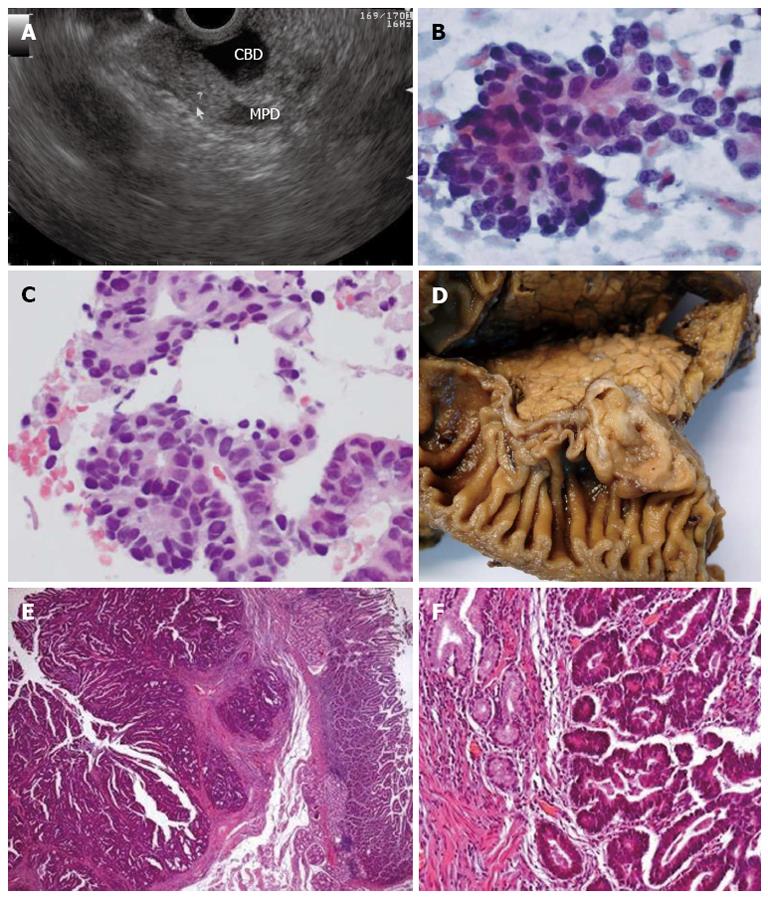
Figure 2 Endoscopic ultrasonography and pathological findings of an ampullary carcinoma.
A: Mildly hyperechoic third-layer lesion adjacent to the ampulla, compressing the bile duct; B, C: Fine-needle aspiration, × 400 magnification; B: Smears with acinar groups, irregularly distributed nuclei, coarse chromatin, conspicuous nucleoli (Papanicolaou); C: Cell-block preparation of aspirated sample [hematoxylin and eosin (HE)]; D: Surgical pathology specimen confirming the full excision of an ampullary carcinoma; E: Ampullary area well-differentiated adenocarcinoma, HE × 25; F: HE × 100.
Figure 3 Endoscopic, endoscopic ultrasonography and pathological findings of hamartomatous polyp.
A: Semipedunculated polyp in the second portion of the duodenum; B: Longitudinal view of the polyp’s stalk-originating from the duodenal wall; C: Top of the polypoid lesion-cross-sectional view; D: Hamartomatous polyp, HE × 25.
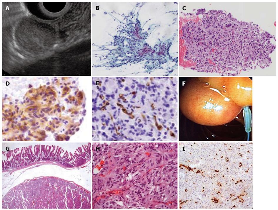
Figure 4 Endoscopic, endoscopic ultrasonography and pathological findings of gangliocytic paraganglioma.
A: Slightly hyperechoic lesion of the third layer of the duodenal wall; B-E: Endoscopic ultrasonography-fine needle aspiration with cytological features suggestive of GIST; B: Few fragments of loose mesenchymal spindle cell tissue fragments (Papanicolaou staining × 100); C: Cell block preparation of aspirated material, discrete nuclear atypia [Hematoxylin and eosin (HE) × 100]; D: Most cells stain positive for CD117 (× 400); E: Rare cells stained with CD34 (× 400); F: Resection of the subepithelial lesion using endoloop; G-I: Histopathological analysis of the resected tumor; G: Duodenal gangliocytic paraganglioma (HE × 25); H: Duodenal gangliocytic paraganglioma (HE × 200); I: Sustentacular S-100 positive cells documented (S100 ×100). GIST: Gastrointestinal stromal tumor.
- Citation: Figueiredo PC, Pinto-Marques P, Mendonça E, Oliveira P, Brito M, Serra D. Duodenal subepithelial hyperechoic lesions of the third layer: Not always a lipoma. World J Gastrointest Endosc 2013; 5(10): 514-518
- URL: https://www.wjgnet.com/1948-5190/full/v5/i10/514.htm
- DOI: https://dx.doi.org/10.4253/wjge.v5.i10.514


