©The Author(s) 2025.
World J Gastrointest Endosc. Sep 16, 2025; 17(9): 110850
Published online Sep 16, 2025. doi: 10.4253/wjge.v17.i9.110850
Published online Sep 16, 2025. doi: 10.4253/wjge.v17.i9.110850
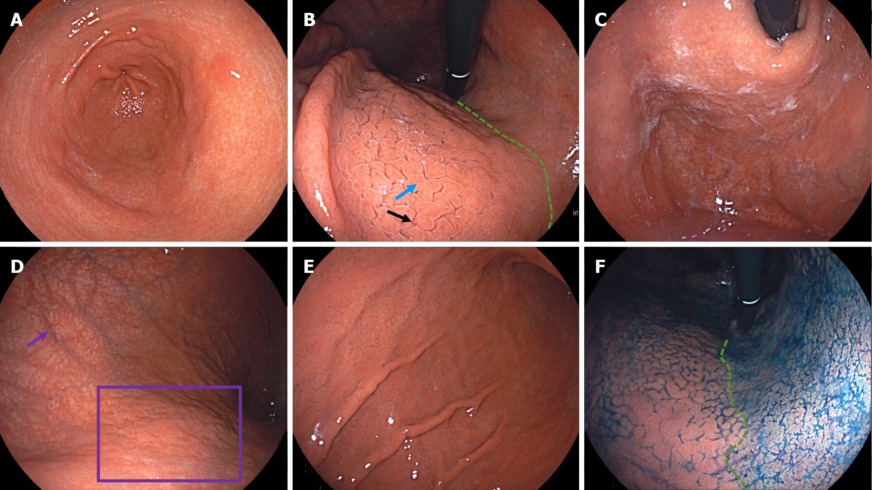
Figure 1 Endoscopic images from the patient in Case 1.
A: Atrophy of the antrum with visible xanthomas and circular wrinkle-like patterns; B: Atrophy along the lesser curvature of the corpus, with a clearly visible atrophic border (green line). Gyrus-like changes on the anterior wall of the corpus in the form of mucosal swelling (blue arrow) and deep fissures (black arrow); C: Adherent white mucus in the fundus; D: Severe atrophy in the upper greater curvature of the corpus, with visible submucosal vessels (purple arrow) after adequate insufflation. Scattered grid-like atrophy (purple box) can be seen on the posterior wall; E: Gyrus-like changes in the mid-to-lower greater curvature of the corpus; F: Indigo carmine staining clearly highlighted the atrophic border.
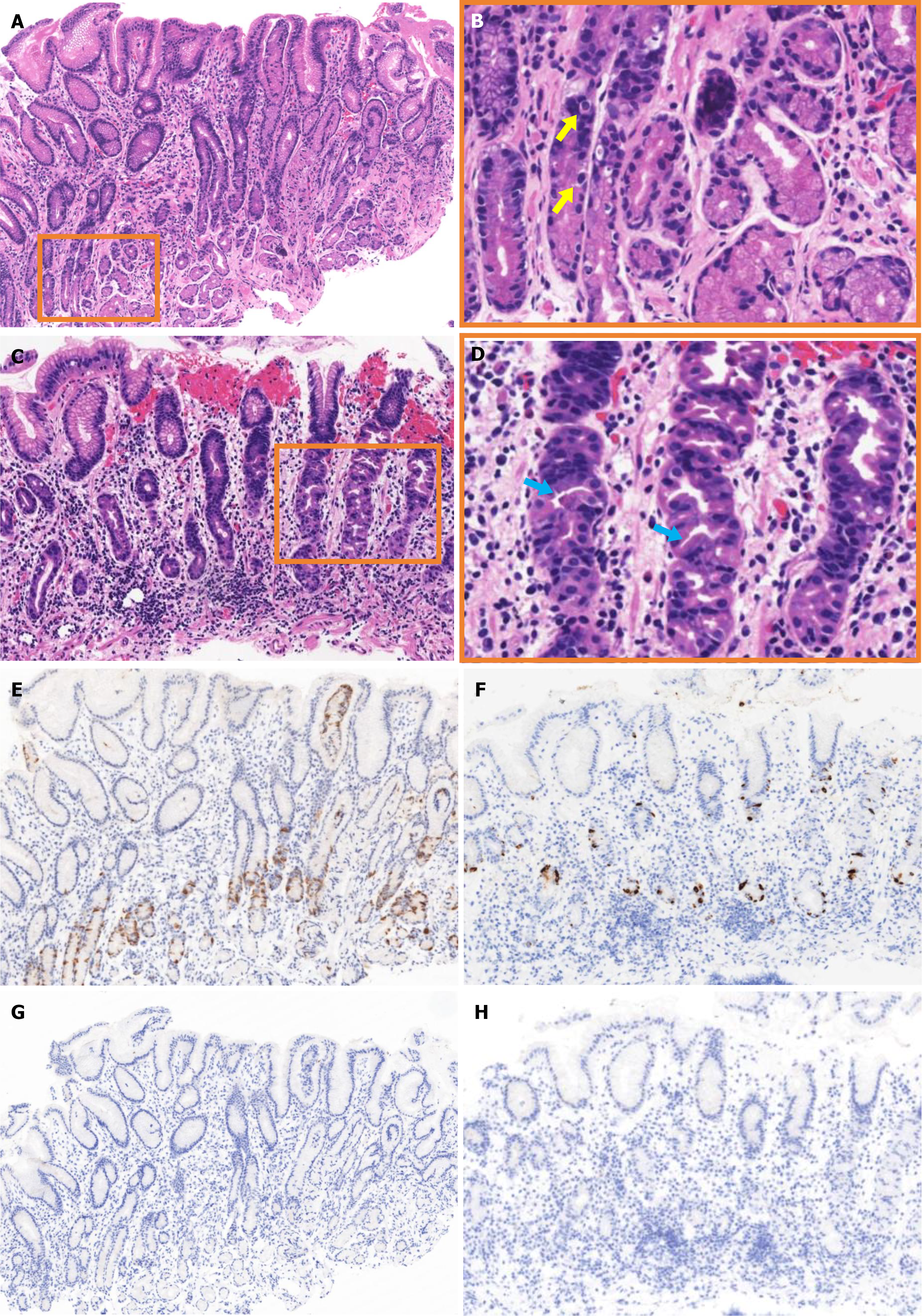
Figure 2 Histopathological images from different regions of the patient’s stomach in Case 1.
A: Hematoxylin and eosin staining of an antral biopsy specimen; B: Magnified view of the boxed area in A (yellow arrows indicate G-cell hyperplasia); C: Hematoxylin and eosin staining of a corporal biopsy specimen; D: Magnified view of the boxed area in C (blue arrows indicate pseudohypertrophy and luminal protrusion of the parietal cells); E: Gastrin staining of an antral biopsy specimen; F: Chromogranin A staining of a corporal biopsy specimen; G: Helicobacter pylori (H. pylori) staining of an antral biopsy specimen; H: H. pylori staining of a corporal biopsy specimen.
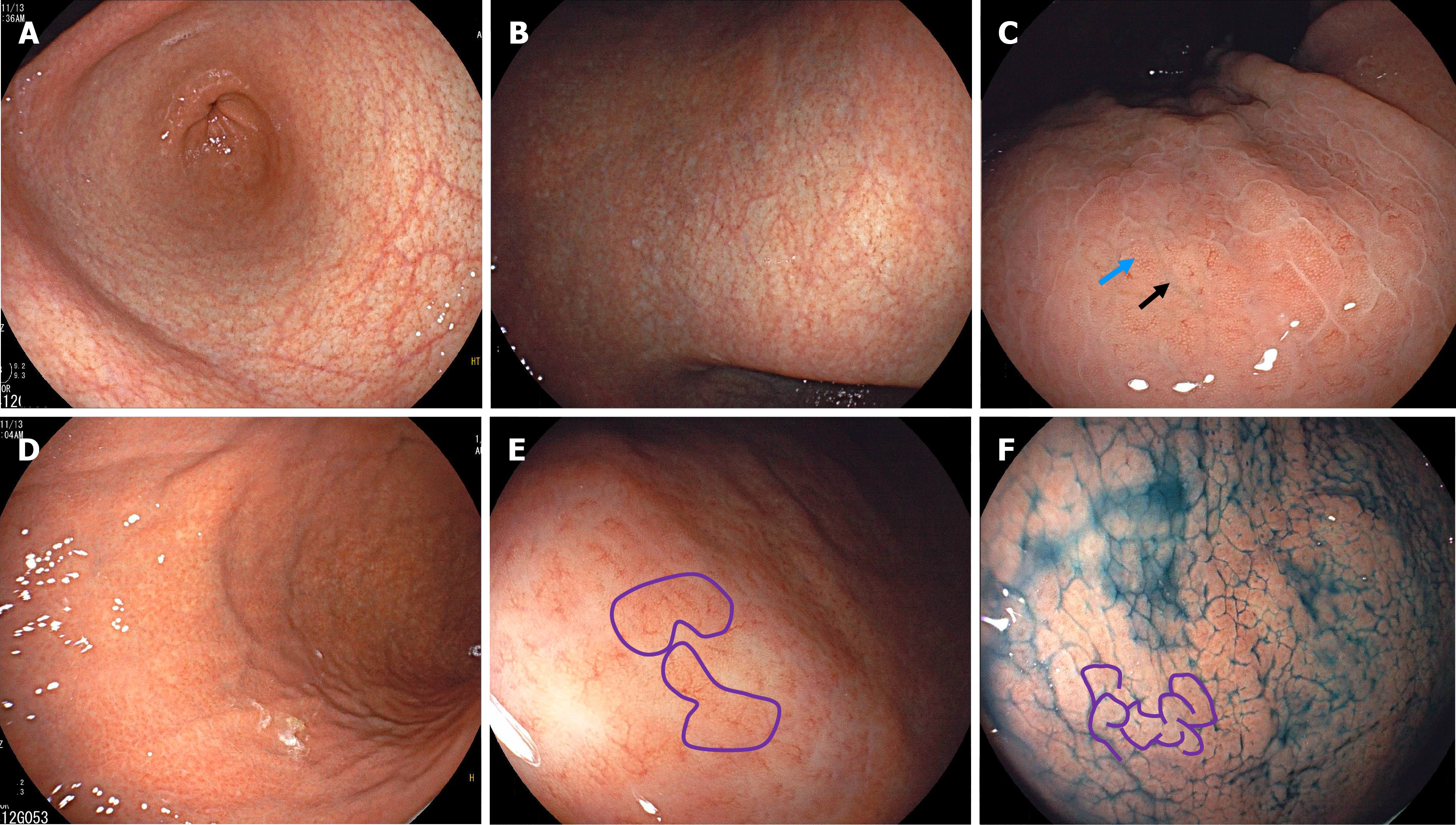
Figure 3 Endoscopic images from the patient in Case 2.
A: Atrophic changes in the antrum; B: Atrophy extending toward the lesser curvature of the corpus; C: A gyrus-like pattern of mild mucosal swelling (blue arrow) and deep fissures (black arrow) can be observed on the anterior wall of the corpus. There were visible collecting venules on the surface of the elevated mucosa; D: Alternating distribution of remnant oxyntic mucosa and atrophic mucosa on the greater curvature of the corpus; E: A close-up view showing remnant oxyntic mucosa with gyrus-like morphology (purple lines) on the greater curvature of the corpus; F: Indigo carmine staining highlighted the widespread gyrus-like changes (purple lines) in the greater curvature of the corpus.
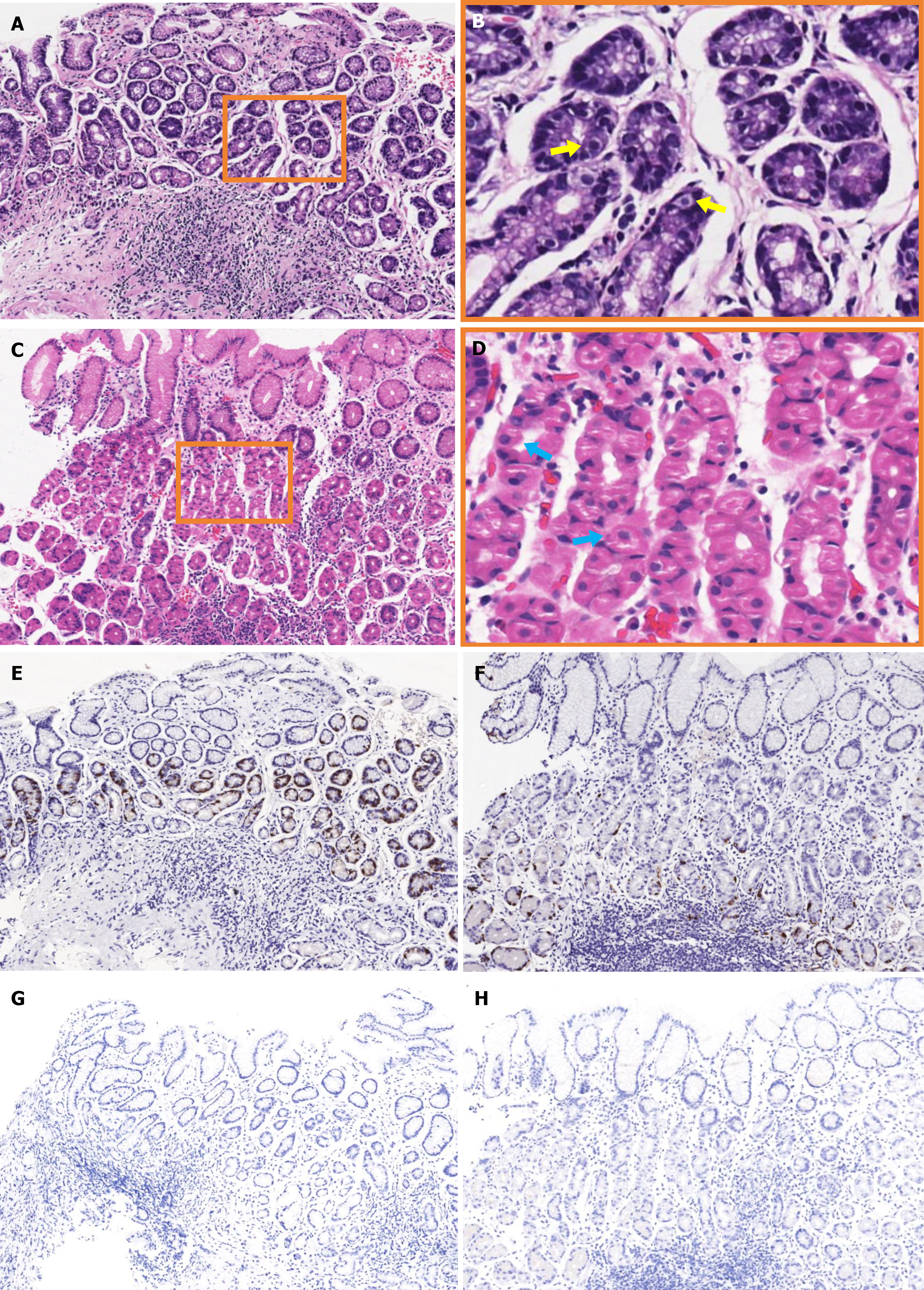
Figure 4 Histopathological images from different regions of the patient’s stomach in Case 2.
A: Hematoxylin and eosin staining of an antral biopsy specimen; B: Magnified view of the boxed area in A (the yellow arrows indicate G-cell hyperplasia); C: Hematoxylin and eosin staining of a corporal biopsy specimen; D: Magnified view of the boxed area in C (blue arrows indicate pseudohypertrophy and luminal protrusion of the parietal cells); E: Gastrin staining of an antral biopsy specimen; F: Chromogranin A staining of a corporal biopsy specimen; G: Helicobacter pylori (H. pylori) staining of an antral biopsy specimen; H: H. pylori staining of a corporal biopsy specimen.
- Citation: Tan CC, Shangguan XL, Lei XM, Deng FF, Wu YP, Zhang GM. Gyrus-like endoscopic changes in autoimmune gastritis with past Helicobacter pylori infection: Two case reports and review of literature. World J Gastrointest Endosc 2025; 17(9): 110850
- URL: https://www.wjgnet.com/1948-5190/full/v17/i9/110850.htm
- DOI: https://dx.doi.org/10.4253/wjge.v17.i9.110850


