©The Author(s) 2025.
World J Gastrointest Endosc. Sep 16, 2025; 17(9): 110424
Published online Sep 16, 2025. doi: 10.4253/wjge.v17.i9.110424
Published online Sep 16, 2025. doi: 10.4253/wjge.v17.i9.110424
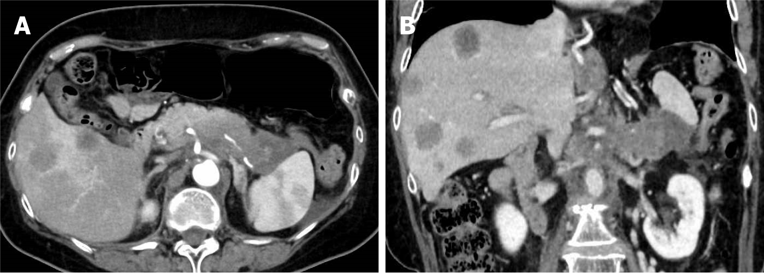
Figure 1 Dynamic computed tomography images of the pancreas at the initial visit to our hospital.
A: Axial view. An irregularly shaped, hypovascular mass is observed in the pancreatic body and tail. Multiple hepatic lesions with low-density cores and enhanced margins are also identified; B: Coronal view. The irregular mass in the pancreatic body and tail directly invades the spleen.
Figure 2 Dynamic computed tomography images of the pancreas at admission for pancreatic fluid leakage.
A: Axial chest view. A large amount of pleural effusion is observed in the left thoracic cavity, along with a small amount in the right thoracic cavity; B: Axial abdominal view; C: Coronal abdominal view. Perisplenic fluid accumulation with partial capsular-like structures is observed.
Figure 3 Findings from the initial endoscopic retrograde cholangiopancreatography.
A: Endoscopic retrograde cholangiopancreatography (ERCP) reveals an irregular stenosis of the main pancreatic duct in the pancreatic body. Distal to the stenosis, the main ductal structure is nearly obliterated by the tumor, and contrast medium is observed leaking into the area of pancreatic fluid leakage through several fine, disrupted ductal structures; B: Guidewire insertion into the leakage site was challenging even with multiple guidewires, but eventually, the guidewire was successfully advanced through an extremely fine tract (indicated by the yellow arrow in Figure 3A), which was not the main contrast-filled route (indicated by the orange arrow in Figure 3A). Attempts to dilate this trans-tumoral passage using a bougie dilator and a balloon dilator were unsuccessful; C: A 5-French endoscopic nasopancreatic drainage tube (Hanaco Medical Co., Saitama, Japan) is placed in the main pancreatic duct proximal to the stenosis.
Figure 4 Findings from the second endoscopic retrograde holangiopancreatography.
A: A guidewire is successfully advanced through the fine tract presumed to be trans-tumoral, reaching the leakage site; B: The tract is dilated using a drill dilator; C: A nasopancreatic drainage tube is placed with its tip positioned in the pancreatic fluid leakage site.
Figure 5 Internalization of the nasopancreatic drainage tube and changes in the pleural effusion volume.
A: An upper gastrointestinal endoscopy is performed to cut the endoscopic nasopancreatic drainage (ENPD) tube within the stomach using a Loop Cutter (Olympus, Tokyo, Japan); B: Chest radiograph at the time of admission. Left pleural effusion is observed; C: Chest radiograph 26 days after ENPD placement. The left pleural effusion is nearly resolved.
- Citation: Makazu M, Koizumi K, Kubota J, Kimura K, Masuda S. Transpapillary drainage of pancreatic fluid leakage via a rigid trans-tumoral tract using a drill dilator: A case report. World J Gastrointest Endosc 2025; 17(9): 110424
- URL: https://www.wjgnet.com/1948-5190/full/v17/i9/110424.htm
- DOI: https://dx.doi.org/10.4253/wjge.v17.i9.110424


