©The Author(s) 2025.
World J Gastrointest Endosc. Jul 16, 2025; 17(7): 107810
Published online Jul 16, 2025. doi: 10.4253/wjge.v17.i7.107810
Published online Jul 16, 2025. doi: 10.4253/wjge.v17.i7.107810
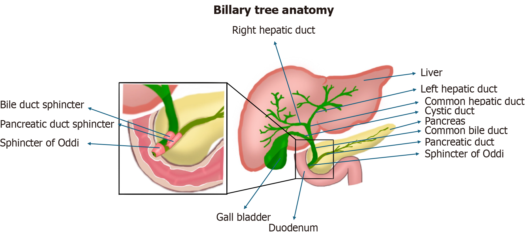
Figure 1
The anatomy of the biliary tract.
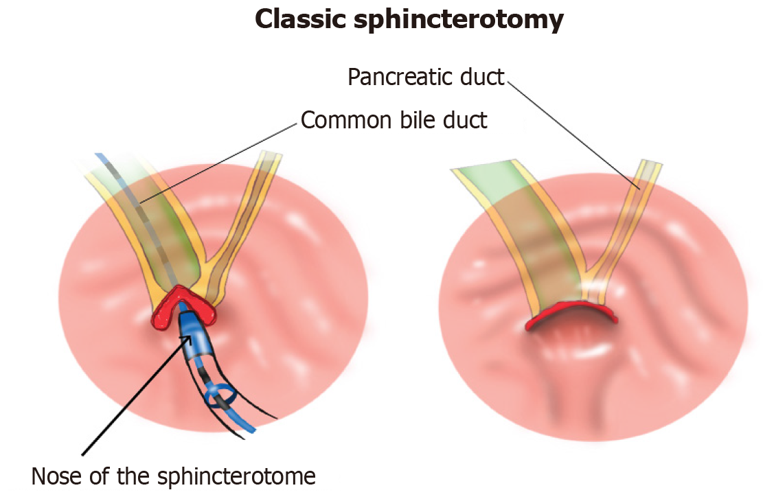
Figure 2
Biliary and pancreatic duct routes in major papilla and classic biliary sphincterotomy.
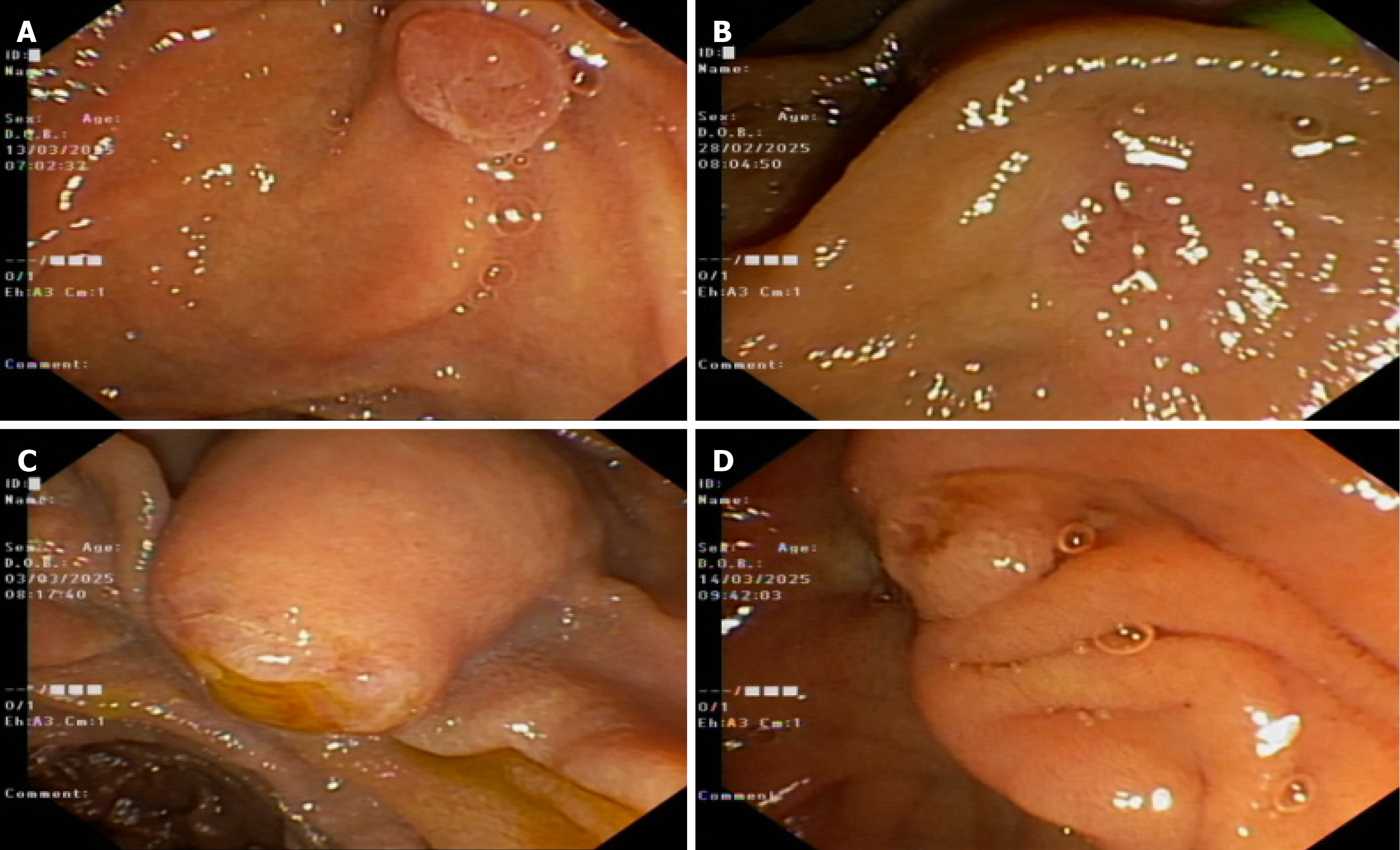
Figure 3 Anatomical variations of major duodenal papilla.
A: Regular; B: Small; C: Protruding; D: Ridged.
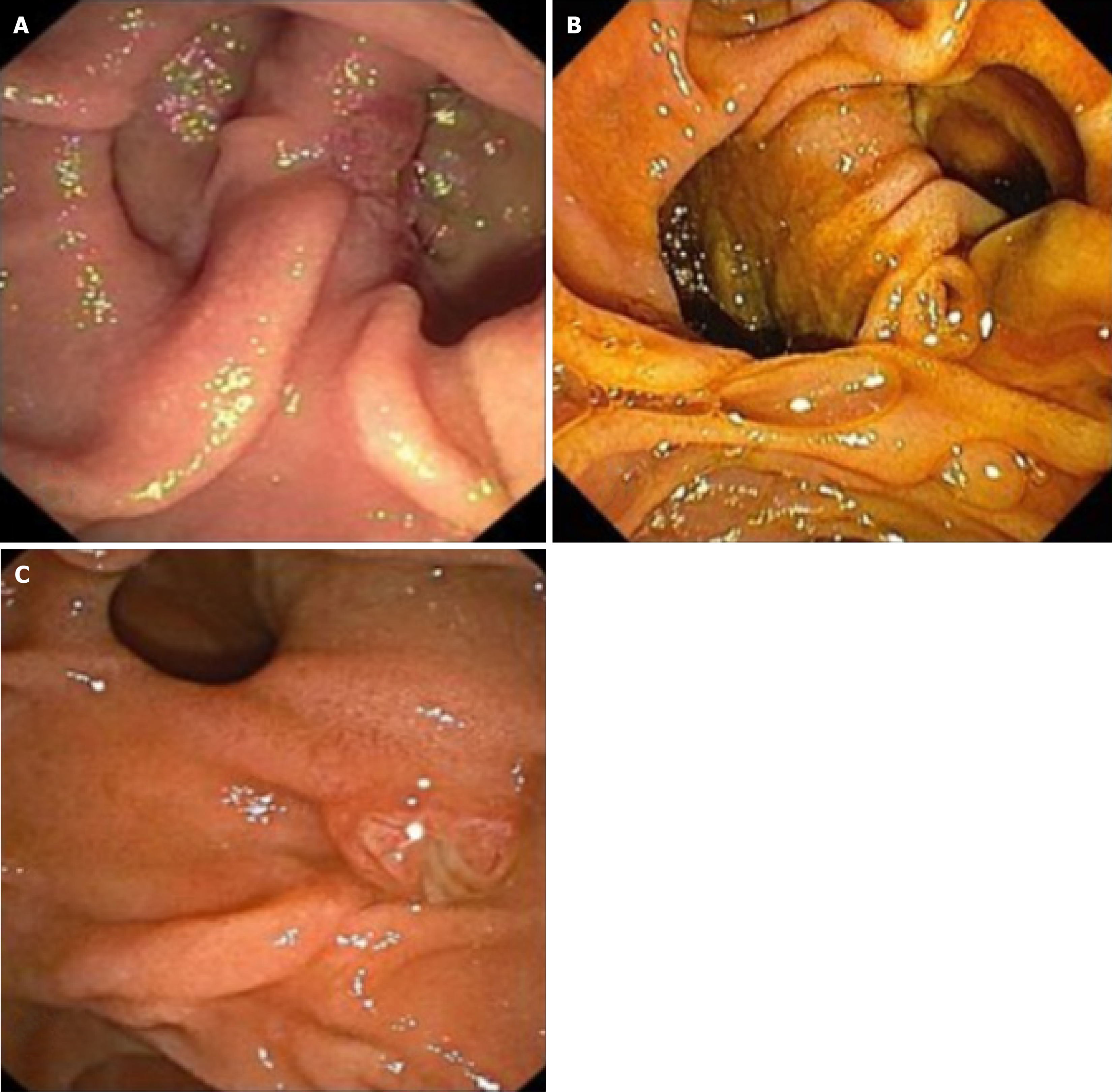
Figure 4 Periampullary diverticula.
A: Type I; B: Type IIa; C: Type III.
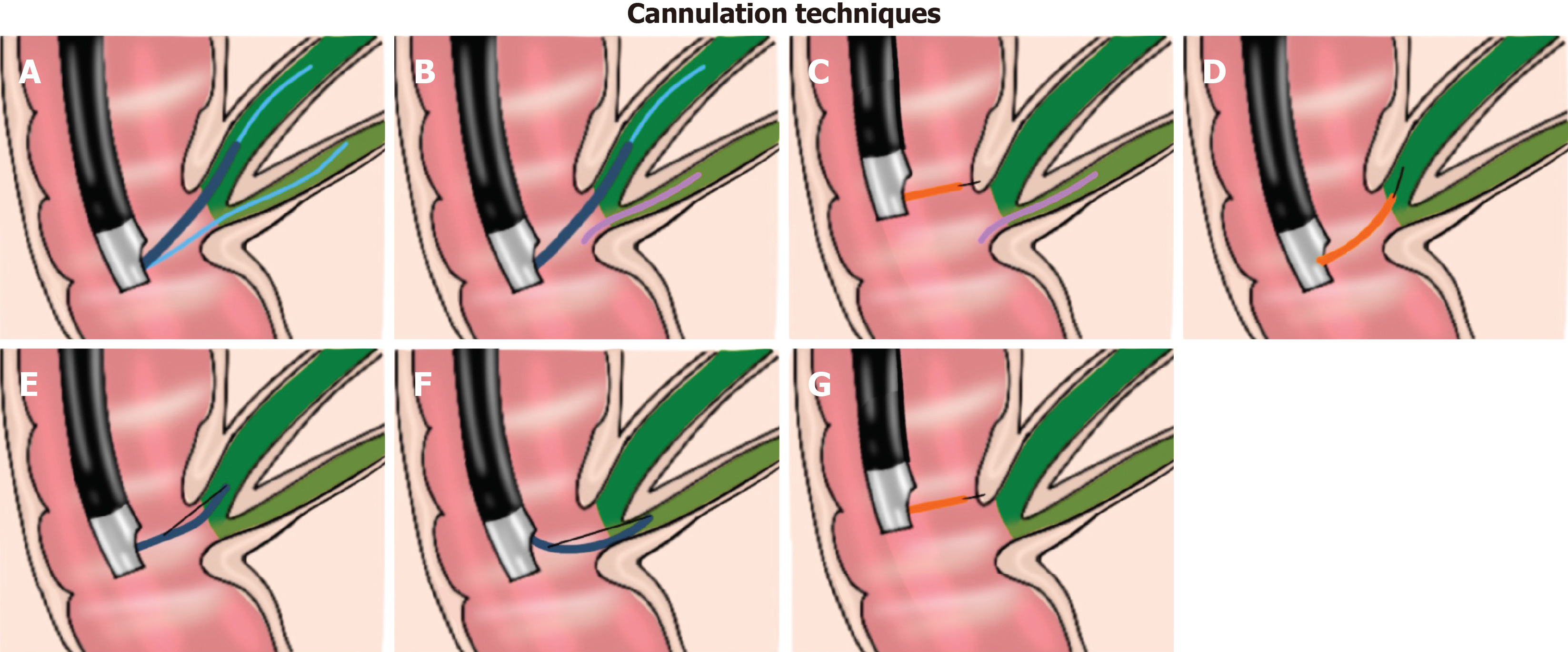
Figure 5 Advanced techniques for biliary cannulation.
A: Double guide-wire; B: Biliary cannulation over pancreatic stenting; C: Precut sphincterotomy over a pancreatic duct stent; D: Precut papillotomy; E: Pull type precut; F: Transpancreatic precut sphincterotomy; G: Precut suprapapillary fistulotomy.
Figure 6
Precut fistulotomy.
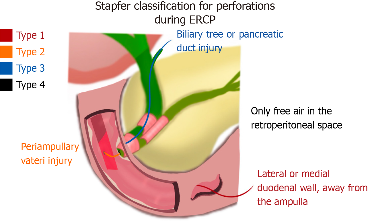
Figure 7 Stapfer classification.
ERCP: Endoscopic retrograde cholangiopancreatography.
- Citation: Ismail A, Abdelwahab MM, Ozercan M, Elnahas O, Bahcecioglu IH, Yalniz M, Tawheed A. Strategies for achieving successful cannulation in endoscopic retrograde cholangiopancreatography: A technical overview. World J Gastrointest Endosc 2025; 17(7): 107810
- URL: https://www.wjgnet.com/1948-5190/full/v17/i7/107810.htm
- DOI: https://dx.doi.org/10.4253/wjge.v17.i7.107810


