Copyright
©The Author(s) 2025.
World J Gastrointest Endosc. Jun 16, 2025; 17(6): 106347
Published online Jun 16, 2025. doi: 10.4253/wjge.v17.i6.106347
Published online Jun 16, 2025. doi: 10.4253/wjge.v17.i6.106347
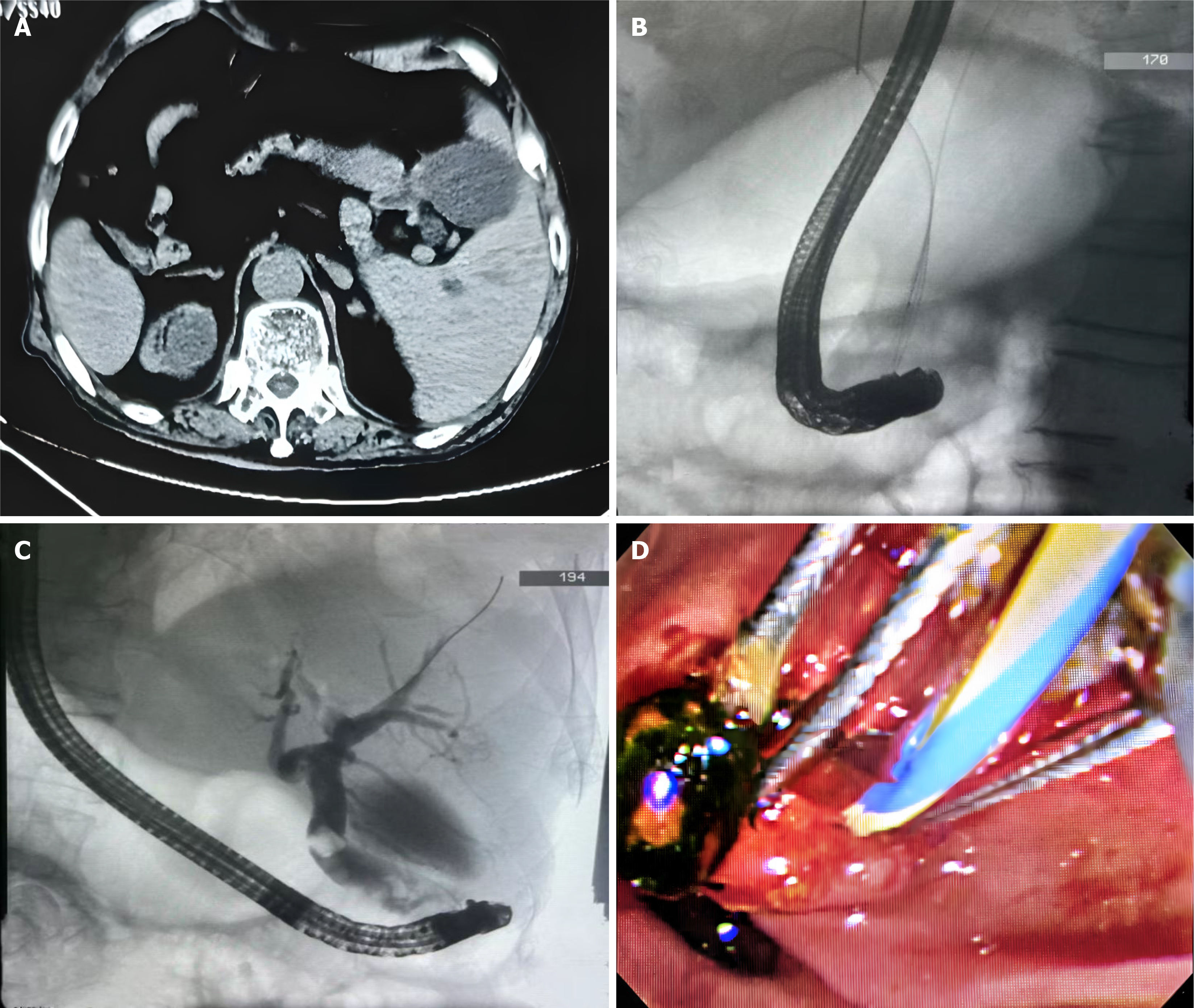
Figure 1 Multimodal imaging and interventional management in situs inversus associated biliary disorders: A pictorial case series.
A: Computed tomography scan showing that the liver and gallbladder are primarily located on the left side of the abdomen, suggesting complete situs inversus viscerum; B: Successful bile duct insertion using the dual-guidewire strategy; C: Cholangiography shows dilated bile ducts and bile duct stones; D: Removed stones visible under endoscopy.
- Citation: Gong KR, Zheng ZL, Li GF, Chen JM. Endoscopic retrograde cholangiopancreatography treatment of cholangitis stone in a patient with total situs inversus: A case report. World J Gastrointest Endosc 2025; 17(6): 106347
- URL: https://www.wjgnet.com/1948-5190/full/v17/i6/106347.htm
- DOI: https://dx.doi.org/10.4253/wjge.v17.i6.106347


