©The Author(s) 2023.
World J Gastrointest Endosc. Dec 16, 2023; 15(12): 745-750
Published online Dec 16, 2023. doi: 10.4253/wjge.v15.i12.745
Published online Dec 16, 2023. doi: 10.4253/wjge.v15.i12.745
Figure 1 Colonoscopy.
A: Distal stenosis; B: Proximal stenosis.
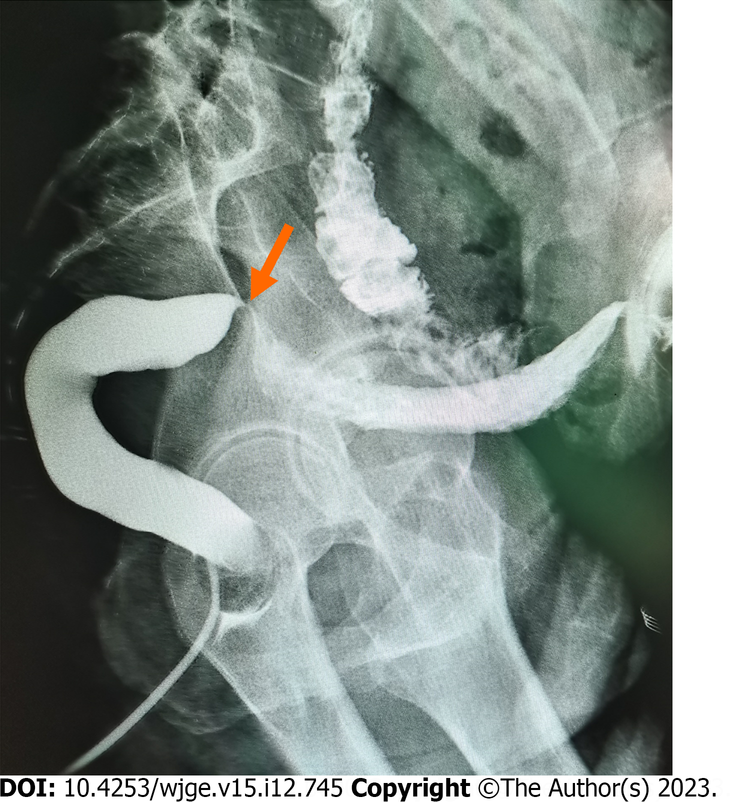
Figure 2
Colonography: Arrow points to the stenosis.
Figure 3 Magnet placement process.
A: The daughter magnet was inserted through the colostomy; B: The parent magnet was inserted through the anus; C: Intraoperative X-ray examination confirmed that the daughter and parent magnets were attracted.
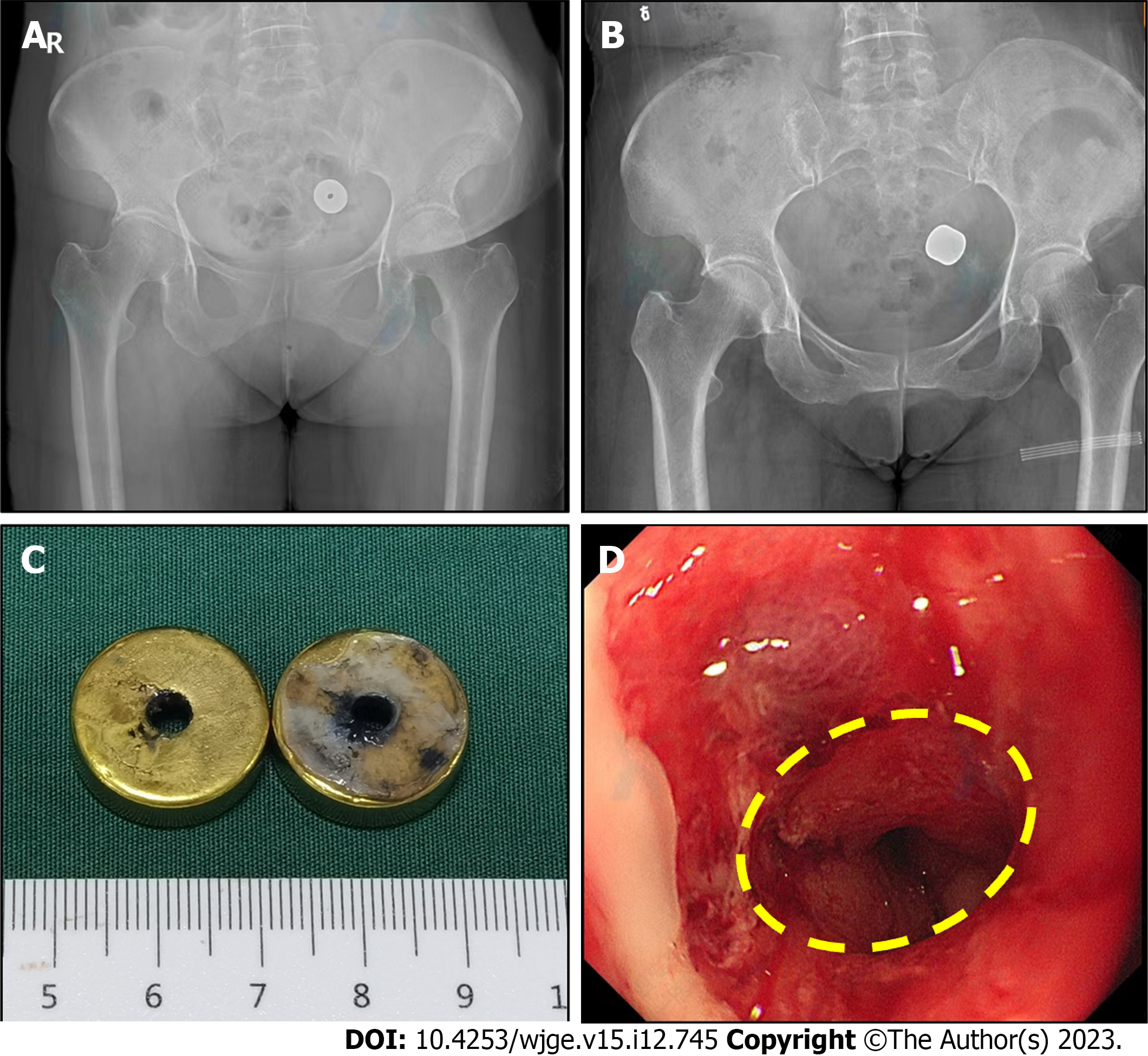
Figure 4 Postoperative X-ray and colonoscopic observations.
A: X-ray examination 1 wk after the surgery; B: X-ray examination 2 wk after the surgery; C: The magnet was removed by colonoscopy 15 d after the surgery; D: The anastomosis was detected on colonoscopy.
- Citation: Zhang MM, Gao Y, Ren XY, Sha HC, Lyu Y, Dong FF, Yan XP. Magnetic compression anastomosis for sigmoid stenosis treatment: A case report. World J Gastrointest Endosc 2023; 15(12): 745-750
- URL: https://www.wjgnet.com/1948-5190/full/v15/i12/745.htm
- DOI: https://dx.doi.org/10.4253/wjge.v15.i12.745


