Copyright
©The Author(s) 2017.
World J Hepatol. Feb 8, 2017; 9(4): 209-216
Published online Feb 8, 2017. doi: 10.4254/wjh.v9.i4.209
Published online Feb 8, 2017. doi: 10.4254/wjh.v9.i4.209
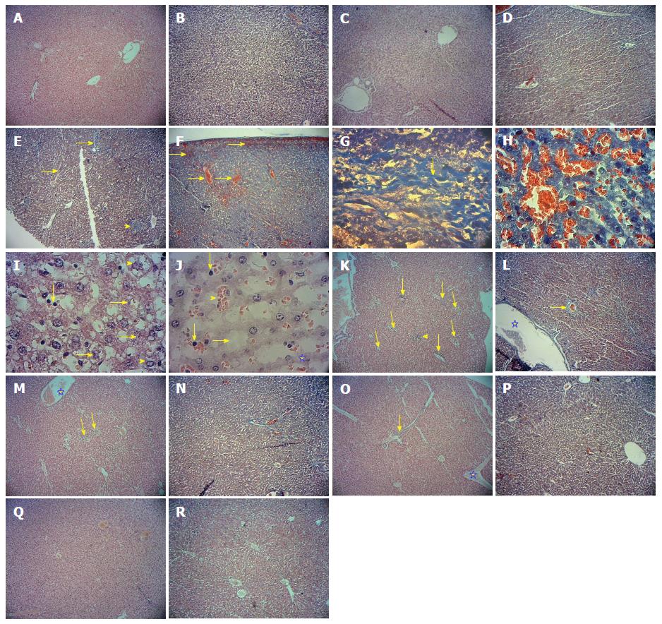
Figure 1 Microscopic views of the liver tissue in study Groups.
Five micron paraffin sections were prepared, stained employing the haematoxylin and eosin stain and histological and pathological changes were studied using a light microscope. A: Control: With a natural structure (Hematoxylin-eosin, 40 × magnification); B: Control: With a natural structure and leukocyte infiltration and congestion cannot be seen (Masson trichrome, 40 × magnification); C: Sham: With a natural structure and Central venous congestion cannot be seen (Hematoxylin-eosin, 40 × magnification); D: Sham: With a natural structure and Collagen fibers cannot be seen. (Masson trichrome, 40 × magnification); E: Paraquat 2 mg/kg: Formation of fibrotic inflamed bridges between liver lobules (thin arrow), the loss of cellular order toward the center (wide arrow), accumulation of collagen fibers and inflammatory cells around the centrilobular vein (arrowhead) (Masson trichrome, 40 × magnification); F: Paraquat 2 mg/kg: Enlarged and congested centrilobular vein (wide arrow), congestion in the sinusoids (thin arrow) (Masson trichrome, 40 × magnification); G: Paraquat 2 mg/kg: Accumulation and progressive of collagen fibers in the liver parenchyma (Masson trichrome, 400 × magnification); H: Paraquat 2 mg/kg: Sever congestion in the sinusoids (Masson trichrome, 400 × magnification); I: Paraquat 2 mg/kg: Sever cellular ballooning (arrowhead), degenerative changes (thin arrow), proliferation and activation of Kupffer cells (wide arrow), (Hematoxylin-eosin, 400 × magnification); J: Paraquat 2 mg/kg: Activation of Kupffer cells (thin arrow), sever congestion in the sinusoids (arrowhead), degenerative changes (asterisk), enlargement of sinusoids spece(wide arrow), (Hematoxylin-eosin, 400X magnification); K: Paraquat + Salep at 40 mg/kg: Infiltration of inflammatory cells around the centrilobular vein (wide arrow), Infiltration of inflammatory cells around the portal space (arrowhead), degenerative changes (thin arrow), (Hematoxylin-eosin, 40 × magnification); L: Paraquat + Salep at 40 mg/kg: Enlargement and congested centrilobular vein (asterisk), accumulation of collagen fibers around the portal space (wide arrow) (Masson trichrome, 40X magnification); M: Paraquat + Salep at 80 mg/kg: Degenerative changes (thin arrow), congested centrilobular vein (asterisk) (Hematoxylin-eosin, 40 × magnification); N: Paraquat + Salep at 80 mg/kg: Decreased infiltration of inflammatory cells around the portal, decreased congestion in the sinusoids and more regular cellular order toward the center (Masson trichrome, 40 × magnification); O: Paraquat + Salep at 160 mg/kg: Infiltration of inflammatory cells around the portal space (wide arrow), congested centrilobular vein (asterisk), (Hematoxylin-eosin, 40 × magnification); P: Paraquat + Salep at 160 mg/kg: More regular cellular order toward the center, more decreased congestion of sinusoids and more decreased infiltration of inflammatory cells in the liver parenchyma (Masson trichrome, 40 × magnification); Q: Paraquat + Salep at 320 mg/kg: Its tissues seem relatively healthy, without any certain pathological changes (Hematoxylin-eosin, 40 × magnification); R: Paraquat + Salep at 320 mg/kg: Its tissues seem relatively healthy, without any certain pathological changes (Masson trichrome, 40 × magnification).
- Citation: Atashpour S, Kargar Jahromi H, Kargar Jahromi Z, Zarei S. Antioxidant effects of aqueous extract of Salep on Paraquat-induced rat liver injury. World J Hepatol 2017; 9(4): 209-216
- URL: https://www.wjgnet.com/1948-5182/full/v9/i4/209.htm
- DOI: https://dx.doi.org/10.4254/wjh.v9.i4.209


