©The Author(s) 2017.
World J Hepatol. Oct 28, 2017; 9(30): 1176-1189
Published online Oct 28, 2017. doi: 10.4254/wjh.v9.i30.1176
Published online Oct 28, 2017. doi: 10.4254/wjh.v9.i30.1176
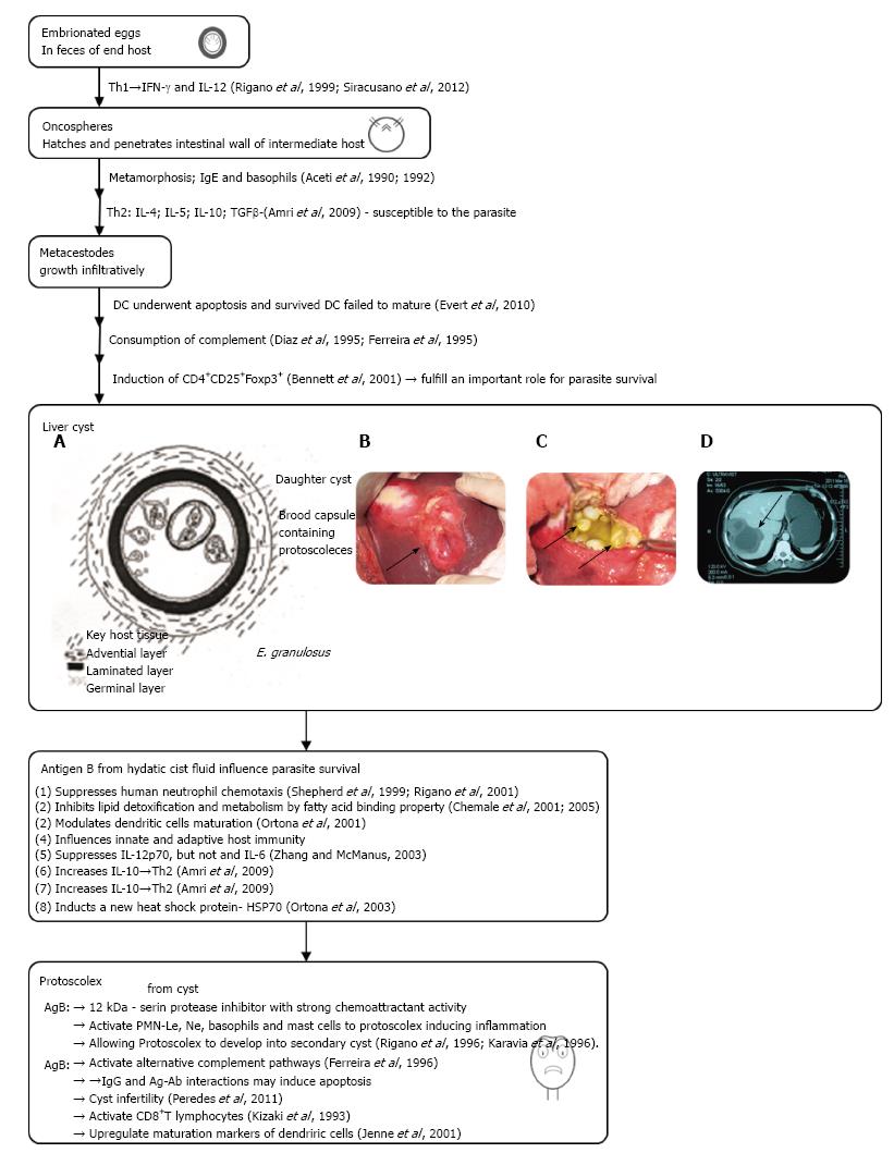
Figure 1 Diagrammatic representation of the development of the Echinococcus granulosus liver cyst.
A, B: Liver cystic echinococcosis, our surgical material; C: Computed tomography imaging of hepatic cystic echinococcosis; D: Abdominal scan of a female patient with a CE3b cyst in the VI liver segment (adapted from Ref. [120]). IL: Interleukin; IFN: Interferon; TGF: Transforming growth factor; PSC: Protoscolece; PMN: Polymorphonuclear.
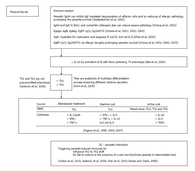
Figure 2 Subversive strategies of Echinococcus granulosus.
DC: Dendritic cell; IL: Interleukin; IFN: Interferon; TGF: Transforming growth factor.
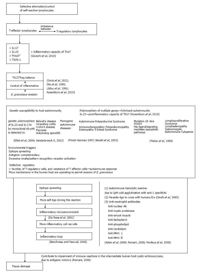
Figure 3 Echinococcus granulosus Induced autoimmunity.
Ags: Antigens.
- Citation: Grubor NM, Jovanova-Nesic KD, Shoenfeld Y. Liver cystic echinococcosis and human host immune and autoimmune follow-up: A review. World J Hepatol 2017; 9(30): 1176-1189
- URL: https://www.wjgnet.com/1948-5182/full/v9/i30/1176.htm
- DOI: https://dx.doi.org/10.4254/wjh.v9.i30.1176


