Copyright
©The Author(s) 2017.
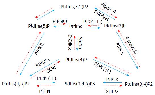
Figure 1 Phosphorylation and dephosphorylation cycles of different phosphoinositides.
Monophosphate, diphosphate and triphosphate phosphoinositides are represented, as are the most widely studied kinases (in blue) and phosphatases (in red) that regulate their expression. PtdIns: Phosphatidylinositol; PI3K: Phosphoinositide 3-kinase; SHIP2: SH2-containing inositol polyphosphate 5-phosphatase; PTEN: Phosphatase and tensin homologue deleted on chromosome 10; OCRL: Inositol polyphosphate 5-phosphatase.
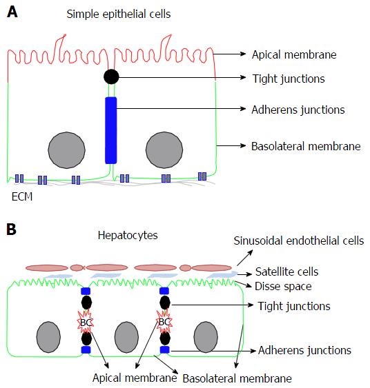
Figure 2 Comparison between simple epithelial cells polarity and hepatocytes polarity.
A: The epithelial cell polarity presents a differentiation of the apical membrane (in red) to the light, and the basolateral membrane (in green) in contact with the extracellular matrix (ECM). Apical and basal membranes are separated with tight junctions (in black) and adherens junctions (in blue); B: The hepatocytes present a more complex polarity, with several apical membranes for the same cell forming bile ducts. The basolateral membrane is in contact with Disse space allowing the exchange with blood vessels.
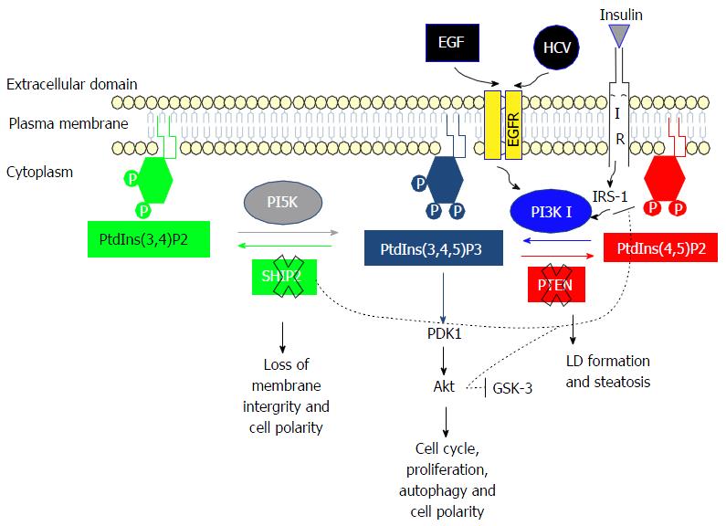
Figure 3 PI3K/Akt pathway and its activation by hepatitis C virus infection.
When EGF binds to EGFR this will activate PI3K I. PI3K activation will increase the level of PtdIns(3,4,5)P3 to phosphorylate Akt and induce cell proliferation and cell polarity. Hepatitis C virus (HCV) is able to bind EGFR at its entry into a cell, thus increasing PI3K activation and the level of PtdIns(3,4,5)P3, the latter being sustained by the down-regulation of SHIP2 and PTEN. The down-regulation of SHIP2 by HCV induces a loss of membrane integrity and cell polarity, while the down regulation of PTEN induces lipid droplet (LD) formation and steatosis. PtdIns: Phosphatidylinositol; PI3K: Phosphoinositide 3-kinase; SHIP2: SH2-containing inositol polyphosphate 5-phosphatase; PTEN: Phosphatase and tensin homologue deleted on chromosome 10; EGFR: Epidermal growth factor receptor.
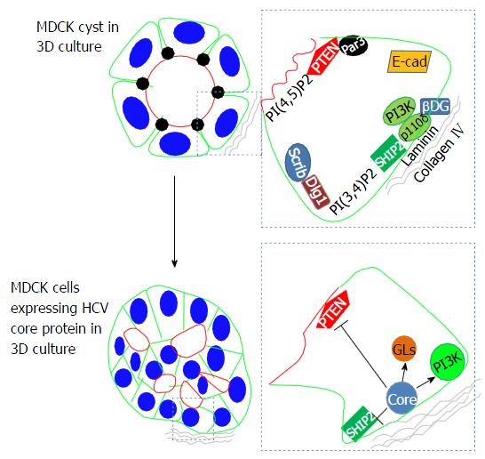
Figure 4 Effects of hepatitis C virus infection on cell polarity.
In a 3D culture, MDCK form a cyst comprising a monolayer of cells around a lumen (the basolateral membrane is indicated in green, the apical membrane in red and the tight junctions in black). The right-hand column shows a zoom of a polarized cell. PI3K is activated by E-cadherin. The p110δ subunit is activated by ECM (Laminin and collagen IV). SHIP2 and PI(3,4)P2 at the basal membrane are responsible for the Dlg1/Scribble complex at the basolateral membrane. PTEN interacts with Par3 at the tight junctions and its lipid product PtdIns(4,5)P2 is localized at the apical membrane. The bottom column represents the multi-lumen phenotype of cysts expressing HCV core protein. The presence of this core protein at the basolateral membrane activates PI3K and inhibits SHIP2 and PTEN, leading to a loss of cell polarity and an accumulation of lipid droplets (LDs). HCV: Hepatitis C virus; PtdIns: Phosphatidylinositol; PI3K: Phosphoinositide 3-kinase; SHIP2: SH2-containing inositol polyphosphate 5-phosphatase; PTEN: Phosphatase and tensin homologue deleted on chromosome 10; MDCK: Madin darby canine kidney; DG: Dystroglycan.
- Citation: Awad A, Gassama-Diagne A. PI3K/SHIP2/PTEN pathway in cell polarity and hepatitis C virus pathogenesis. World J Hepatol 2017; 9(1): 18-29
- URL: https://www.wjgnet.com/1948-5182/full/v9/i1/18.htm
- DOI: https://dx.doi.org/10.4254/wjh.v9.i1.18


