Copyright
©The Author(s) 2016.
World J Hepatol. Mar 28, 2016; 8(9): 446-451
Published online Mar 28, 2016. doi: 10.4254/wjh.v8.i9.446
Published online Mar 28, 2016. doi: 10.4254/wjh.v8.i9.446
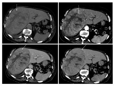
Figure 1 Contrast enhanced computed tomography images of hepatocellular carcinoma.
A 55 years old male, diabetic, presented with upper abdominal pain (arrows shows the lesion in different phases with clear washout at the venous phase).
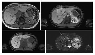
Figure 2 Contrast enhanced magnetic resonance images of focal nodular hyperplasia.
A 30 years old female, medically free, had abdominal pain; ultrasonography showed gallstones and liver lesion (arrows shows the lesion with the characteristic central scar of FNH).
- Citation: Algarni AA, Alshuhri AH, Alonazi MM, Mourad MM, Bramhall SR. Focal liver lesions found incidentally. World J Hepatol 2016; 8(9): 446-451
- URL: https://www.wjgnet.com/1948-5182/full/v8/i9/446.htm
- DOI: https://dx.doi.org/10.4254/wjh.v8.i9.446


