Copyright
©The Author(s) 2016.
World J Hepatol. May 28, 2016; 8(15): 659-664
Published online May 28, 2016. doi: 10.4254/wjh.v8.i15.659
Published online May 28, 2016. doi: 10.4254/wjh.v8.i15.659
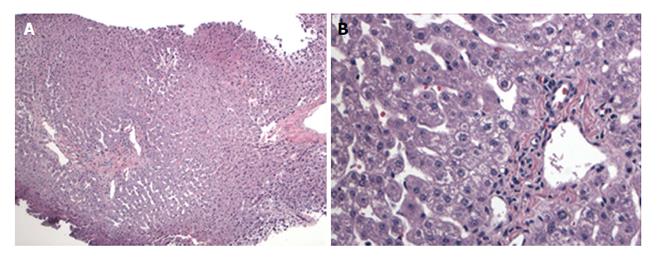
Figure 1 Biopsy of the donor liver showing a lack of obvious pathologic changes.
Specifically there is no steatosis, fibrosis or inflammation (H and E stain). A: Low magnification (× 100); B: Higher magnification (× 200) of the biopsy.
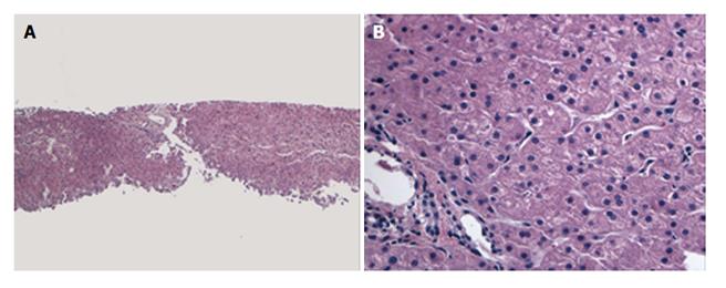
Figure 2 Biopsy of liver graft from case 1 performed 25 mo later showing a lack of significant pathologic changes.
Specifically, no steatosis is noted (H and E stain). A: Low magnification (× 100); B: Higher magnification (× 200) of the biopsy.
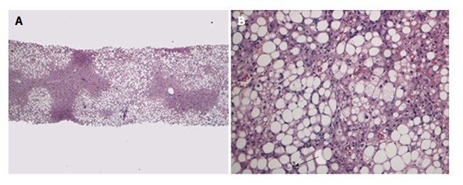
Figure 3 Biopsy of the liver graft from case 2 about 3 years post split liver transplantation showing marked macrovesicular steatosis (H and E stain).
A: Low magnification (× 100); B: Higher magnification showing rare Mallory Denk bodies (arrow), ballooned hepatocytes and mild lobular inflammation (× 200). Trichrome stain revealed mild sinusoidal fibrosis (not shown here).
- Citation: Boga S, Munoz-Abraham AS, Rodriguez-Davalos MI, Emre SH, Jain D, Schilsky ML. Host factors are dominant in the development of post-liver transplant non-alcoholic steatohepatitis. World J Hepatol 2016; 8(15): 659-664
- URL: https://www.wjgnet.com/1948-5182/full/v8/i15/659.htm
- DOI: https://dx.doi.org/10.4254/wjh.v8.i15.659


