Copyright
©The Author(s) 2015.
World J Hepatol. Dec 28, 2015; 7(30): 2933-2939
Published online Dec 28, 2015. doi: 10.4254/wjh.v7.i30.2933
Published online Dec 28, 2015. doi: 10.4254/wjh.v7.i30.2933
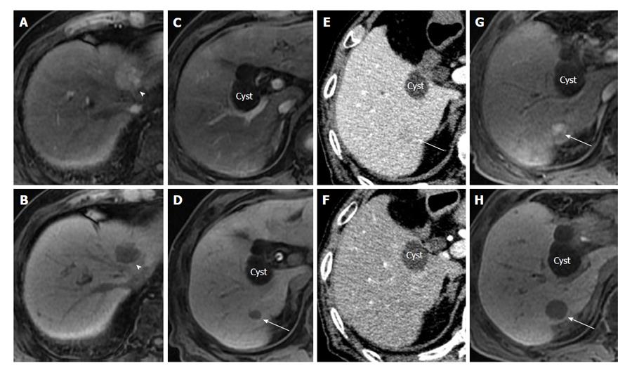
Figure 1 An 81-year-old man with hypervascular (progressed) hepatocellular carcinoma and early hepatocellular carcinoma.
Preoperative EOB-MRI showed hypervascular HCC (arrowhead), 2.6 cm in diameter, in the medial section of the liver. The nodule showed hypervascularity in the hepatic arterial-dominant phase (A) and hypointensity in the hepatobiliary phase (B) of EOB-MRI. Preoperative EOB-MRI also showed a small hypointense nodule (arrow), 0.9 cm in diameter, in the posterior section of the liver in the hepatobiliary phase (D). This nodule showed no arterial enhancement in the hepatic arterial-dominant phase of EOB-MRI (C) or CTHA (F), but showed slight hypoattenuation on CTAP (E). This patient underwent left hepatic resection for hypervascular HCC. EOB-MRI taken 5 mo after hepatic resection showed an enlarged hypointense nodule in the posterior section (H) and hypervascularity of the nodule in the hepatic arterial-dominant phase (G). The recurrence of hypervascular HCC that originated from residual early HCC after hepatic resection was demonstrated. HCC: Hepatocellular carcinoma; EOB-MRI: Gadoxetic acid- or gadolinium ethoxybenzyl diethylenetriamine pentaacetic acid-enhanced magnetic resonance imaging; CTHA: Computed tomography during hepatic arteriography; CTAP: Computed tomography arterioportography.
- Citation: Matsuda M. Clinical value of gadoxetic acid-enhanced magnetic resonance imaging in surgery for hepatocellular carcinoma - with a special emphasis on early hepatocellular carcinoma. World J Hepatol 2015; 7(30): 2933-2939
- URL: https://www.wjgnet.com/1948-5182/full/v7/i30/2933.htm
- DOI: https://dx.doi.org/10.4254/wjh.v7.i30.2933


