Copyright
©2013 Baishideng Publishing Group Co.
World J Hepatol. Sep 27, 2013; 5(9): 487-495
Published online Sep 27, 2013. doi: 10.4254/wjh.v5.i9.487
Published online Sep 27, 2013. doi: 10.4254/wjh.v5.i9.487
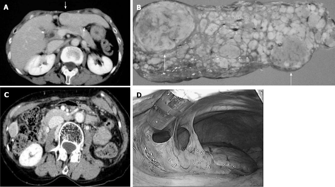
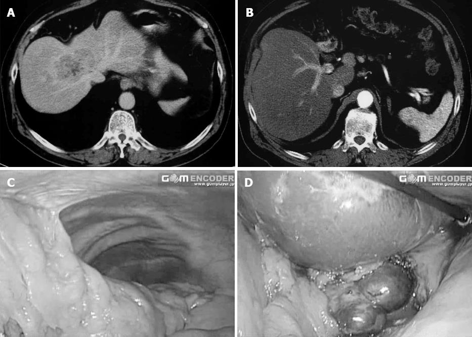
Figure 1 Repeat pure laparoscopic hepatectomy for patients with liver cirrhosis and hepatocellular carcinomas was feasible and safe: case 1.
A: Computed tomorgraphy scan shows two hepatocellular carcinomas (HCC) in segment 3; B: The tumors (arrows) resected laparoscopycally.; C: A 69-year-old woman with type-C liver cirrhosis developed a new HCC on the caudal edge of segment 6 of the liver 2 years after the first hepatectomy; D: At the second laparoscopic hepatectomy, there was only mild adhesion around the resected area
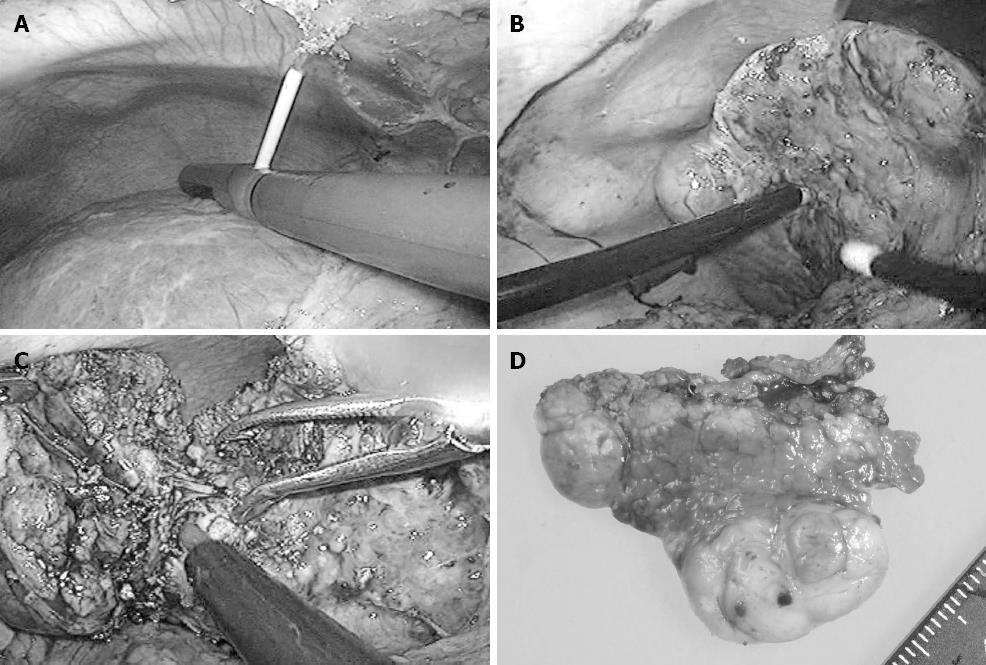
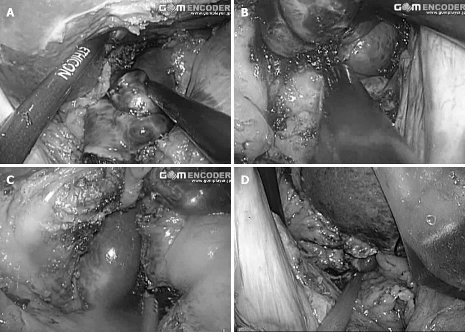
Figure 2 Repeat pure laparoscopic hepatectomy for patients with liver cirrhosis and hepatocellular carcinomas was feasible and safe: case 1.
The patient also had two early lesions in segment four, which was treated with laparoscopic microwave coagulation therapy (A). After ablation therapy, the hepatocellular carcinomas (HCC) in segment 6 (B) was resected laparoscopically (C). The resected specimen (D) showed a single nodular HCC. The patient also underwent third hepatectomy for the lesion in segment one next to right adrenal gland two years after this operation.
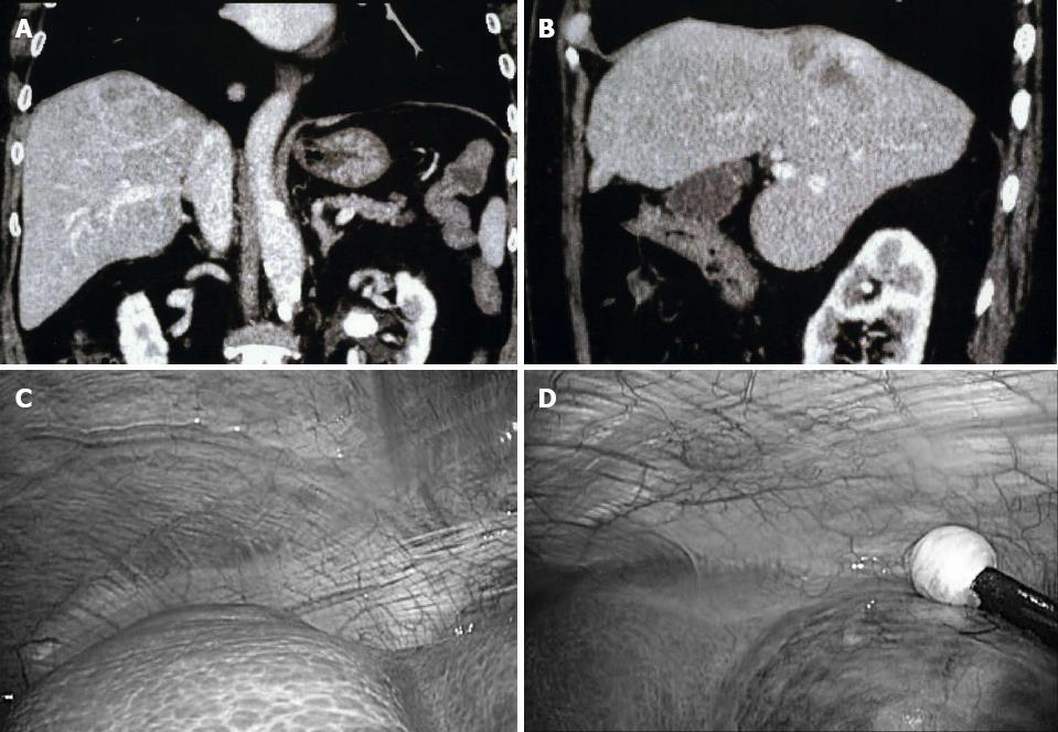
Figure 3 Pure laparoscopic hepatectomy is efficient in the subphrenic space: case 2.
An 80-year-old woman with liver cirrhosis developed a hepatocellular carcinomas in the dorsal area of subsegment 8c of the liver revealed in computed tomography examination (A and B). Since the tumor compressed the right hepatic vein and her liver function seemed not to tolerate right hepatectomy or extended anterior sectionectomy, she underwent partial resection of the liver with the dissection and exposure of right hepatic vein and tumor capsule in pure laparoscopic hepatectomy. The tumor was located deeply in the subphrenic space (C) just next to the attachment of retro-peritoneum (D).
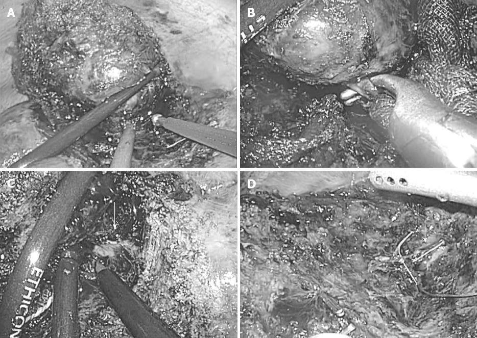
Figure 4 Pure laparoscopic hepatectomy is efficient in the subphrenic space: case 2.
A: Resection of the tumor with the exposure of the capsule; B: Encircling and dividing of the direct branch of the right hepatic vein; C: Exposure of right hepatic vein; D: Cutting surface after resection.
Figure 5 Pure laparoscopic hepatectomy is efficient between the adhesions and the peri-inferior vena cava area: case 3.
Two years after a central bisectionectomy for hepatocellular carcinomas (HCC) at the roots of hepatic veins (A), a 66-year-old man developed a new prominent HCC on the left caudate lobe of the liver (B). Following the second pure laparoscopic hepatectomy, there was massive adhesion in the area of right upper abdomen (C). However, good view and access to the tumor were obtained with the dissection of omentum minus (D).
Figure 6 Pure laparoscopic hepatectomy is efficient between the adhesions and the peri-inferior vena cava area: case 3.
A and B: Resection of the tumor; C and D: The view after the resection of the tumor.
- Citation: Morise Z, Kawabe N, Kawase J, Tomishige H, Nagata H, Ohshima H, Arakawa S, Yoshida R, Isetani M. Pure laparoscopic hepatectomy for hepatocellular carcinoma with chronic liver disease. World J Hepatol 2013; 5(9): 487-495
- URL: https://www.wjgnet.com/1948-5182/full/v5/i9/487.htm
- DOI: https://dx.doi.org/10.4254/wjh.v5.i9.487


