©2013 Baishideng Publishing Group Co.
World J Hepatol. Apr 27, 2013; 5(4): 230-233
Published online Apr 27, 2013. doi: 10.4254/wjh.v5.i4.230
Published online Apr 27, 2013. doi: 10.4254/wjh.v5.i4.230
Figure 1 Physical examination.
A: The skin lesions in the forehead and upper eyelids; B: The skin lesions in the neck; C: Erythema to the nail fold.
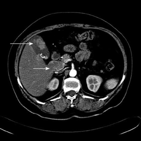
Figure 2 Abdominal computed tomography.
Gallbladder carcinoma (long arrow) with lymph node metastasis (short arrow).
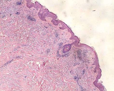
Figure 3 Biopsy of skin.
Mild degree of hyperkeratosis, hair follicle angle plug, epidermal atrophy, basal cell liquefaction degeneration, dermal papilla edema, agglomerate lymphocytic infiltrate around vessels, and dermal adnexa.
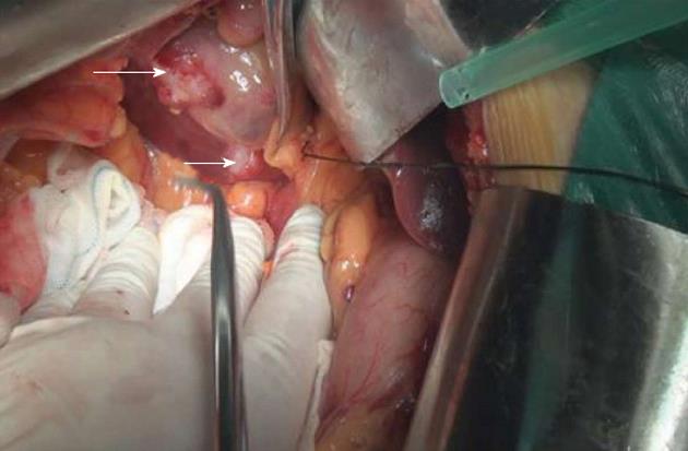
Figure 4 Radical resection of the gallbladder was performed.
A hard, whitish mass located at the fundus of gallbladder (long arrow) and a hard, pale lump was seen in the liver surface (short arrow), between the gallbladder and the inferior vena cava.
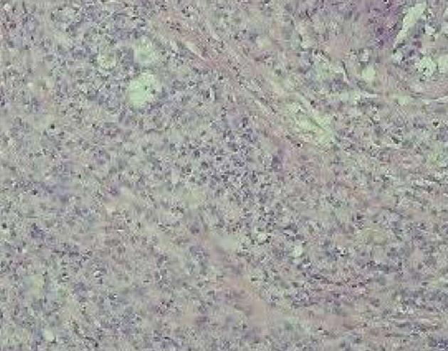
Figure 5 Histopathology of tumor.
Low differentiated gallbladder adenocarcinoma, involving all layers of the gallbladder and infringing upon nerves.
- Citation: Ni QF, Liu GQ, Pu LY, Kong LL, Kong LB. Dermatomyositis associated with gallbladder carcinoma: A case report. World J Hepatol 2013; 5(4): 230-233
- URL: https://www.wjgnet.com/1948-5182/full/v5/i4/230.htm
- DOI: https://dx.doi.org/10.4254/wjh.v5.i4.230


