Copyright
©2012 Baishideng Publishing Group Co.
World J Hepatol. Dec 27, 2012; 4(12): 374-381
Published online Dec 27, 2012. doi: 10.4254/wjh.v4.i12.374
Published online Dec 27, 2012. doi: 10.4254/wjh.v4.i12.374
Figure 1 Classification of B-mode ultrasonographic images of small hepatocellular carcinoma.
A-D: Hepatocellular carcinoma nodules < 3 cm in diameter were classified into two groups using B-mode ultrasonography: Type 1 with halo (A) and type 2 without halo. Type 2 was then further categorized into three subgroups: Type 2a, homogenous hyperechoic (B); Type 2b, hypoechoic with smooth margins (C); Type 2c, hypoechoic with irregular or unclear margins (D). Hepatocellular carcinoma nodules are indicated by arrows.
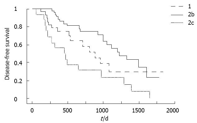
Figure 2 Recurrence-free survival curves according to B-mode ultrasound classification.
Recurrence-free survival was significantly shorter for type 2c hepatocellular carcinoma than for other types. P = 0.0454 type 1 vs type 2c; P = 0.0005 type 2b vs type 2c.
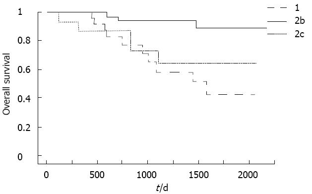
Figure 3 Survival curves according to B-mode ultrasound classification.
Survival was significantly longer for type 2b than for other types. P = 0.0006 type 1 vs type 2b; P = 0.0165 type 2b vs type 2c; P = 0.4473 type 1 vs type 2c.
- Citation: Moribata K, Tamai H, Shingaki N, Mori Y, Shiraki T, Enomoto S, Deguchi H, Ueda K, Inoue I, Maekita T, Iguchi M, Ichinose M. Ultrasonogram of hepatocellular carcinoma is associated with outcome after radiofrequency ablation. World J Hepatol 2012; 4(12): 374-381
- URL: https://www.wjgnet.com/1948-5182/full/v4/i12/374.htm
- DOI: https://dx.doi.org/10.4254/wjh.v4.i12.374


