Copyright
©2012 Baishideng Publishing Group Co.
World J Hepatol. Oct 27, 2012; 4(10): 288-290
Published online Oct 27, 2012. doi: 10.4254/wjh.v4.i10.288
Published online Oct 27, 2012. doi: 10.4254/wjh.v4.i10.288
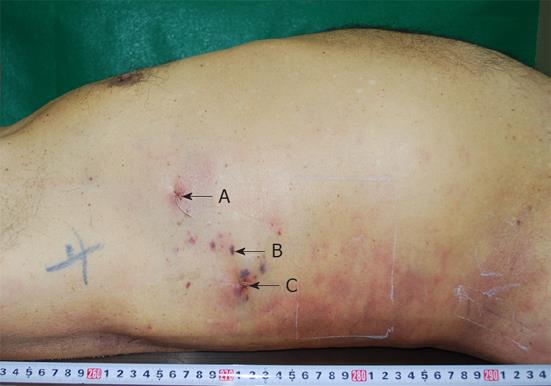
Figure 1 Three centesis scars on the right side of the chest.
As shown in the figure, ventral, middle, and dorsal centeses were assigned as (A), (B) and (C), respectively.
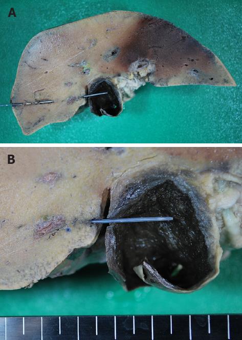
Figure 2 Frontal section of liver along pathway (C).
A: Pathway (C) reaches the gallbladder transversely through the right hepatic lobe; B: Magnified photograph around the gallbladder. A small space between the liver and gallbladder is revealed, and the percutaneous transhepatic gallbladder drainage pathway is partially exposed into the peritoneal cavity. The pathway crosses the intrahepatic connective tissue area near the liver surface.
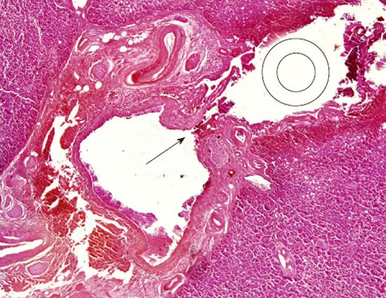
Figure 3 Microscopic specimen at the crossover site of intrahepatic connective tissue and the percutaneous transhepatic gallbladder drainage pathway (hematoxylin-eosin staining).
There is a rupture of the arterial wall near the catheter pathway and intrahepatic bleeding along the percutaneous transhepatic gallbladder drainage pathway. The arrow dead shows the point where the intrahepatic artery is ruptured, and the double circle shows a cross section of the catheter on the pathway (C).
- Citation: Ihama Y, Fukazawa M, Ninomiya K, Nagai T, Fuke C, Miyazaki T. Peritoneal bleeding due to percutaneous transhepatic gallbladder drainage: An autopsy report. World J Hepatol 2012; 4(10): 288-290
- URL: https://www.wjgnet.com/1948-5182/full/v4/i10/288.htm
- DOI: https://dx.doi.org/10.4254/wjh.v4.i10.288


