©2010 Baishideng Publishing Group Co.
World J Hepatol. Sep 27, 2010; 2(9): 362-366
Published online Sep 27, 2010. doi: 10.4254/wjh.v2.i9.362
Published online Sep 27, 2010. doi: 10.4254/wjh.v2.i9.362
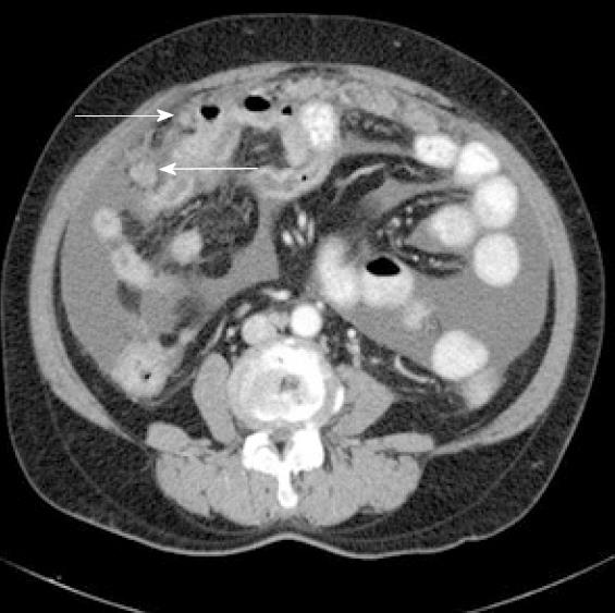
Figure 1 Abdominopelvic computed tomography scan showing ascites and contrast-enhanced peritoneal nodules.
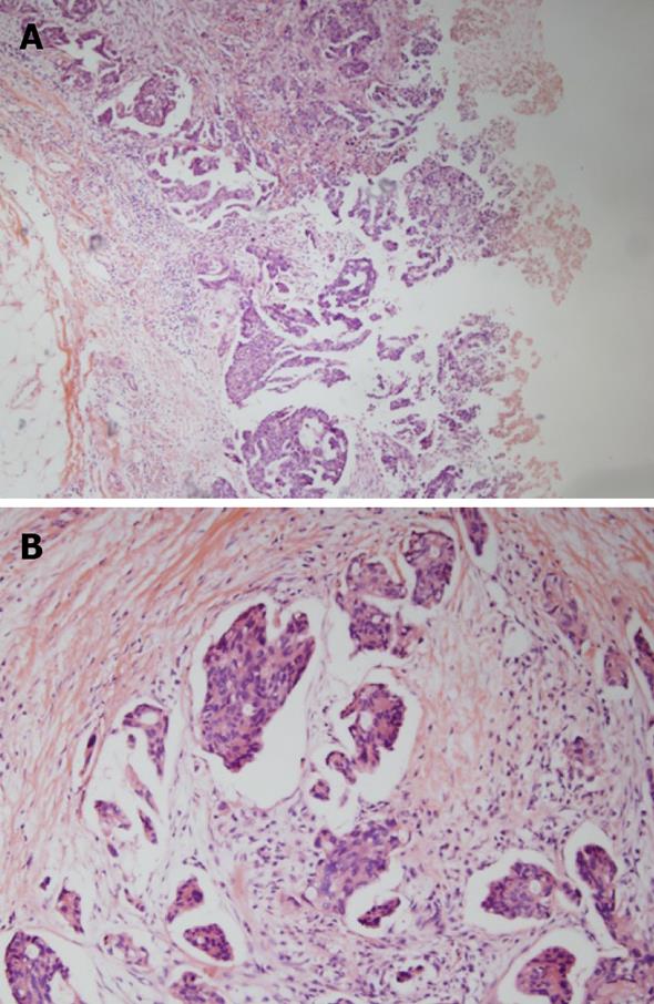
Figure 2 Microscopic findings of the peritoneal nodules revealed papillary serous adenocarcinoma with infiltrative papillary clusters (A, H&E stain × 40) and tumor nests with characteristic retraction artifacts in the stroma (B, H&E stain × 100).
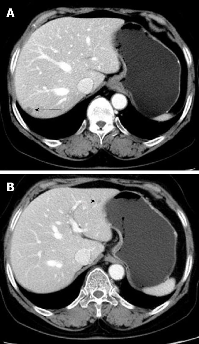
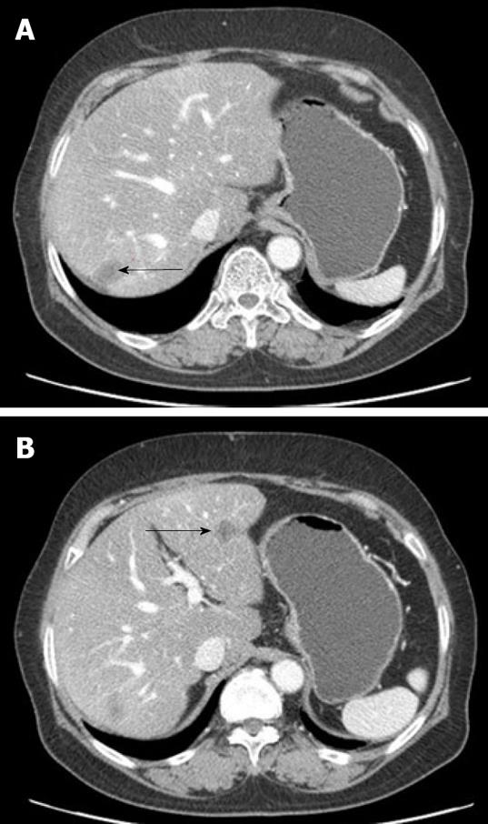
Figure 3 Computed tomography scan showing two hepatic nodules with peripheral enhancement (arrows).
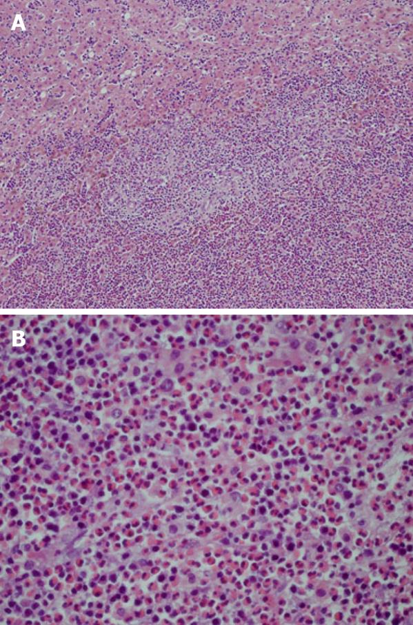
Figure 4 Liver biopsy specimens revealed an inflammatory cell infiltration among the hepatocytes and sinusoids, and partial abscess formation (A, H&E stain × 100).
The inflammatory infiltrate was composed primarily of eosinophils (B, H&E stain × 400).
Figure 5 Computed tomography scan showing two recurrent lesions around the previous operative sites (arrows).
Figure 6 Pathological findings of the specimens from the secondary operation showed chronic inflammation with necrosis.
A: A well-demarcated inflammatory nodule was identified within the liver parenchyma (H&E stain × 40); B: Lymphoplasma cell infiltration was also noted in the peripheral portion of the nodule (H&E stain × 100); C: The nodule was filled with inflammatory necrotic debris (H&E stain × 200).
- Citation: Shim HJ. Tertiary syphilis mimicking hepatic metastases of underlying primary peritoneal serous carcinoma. World J Hepatol 2010; 2(9): 362-366
- URL: https://www.wjgnet.com/1948-5182/full/v2/i9/362.htm
- DOI: https://dx.doi.org/10.4254/wjh.v2.i9.362


