©2010 Baishideng.
World J Hepatol. Jun 27, 2010; 2(6): 208-220
Published online Jun 27, 2010. doi: 10.4254/wjh.v2.i6.208
Published online Jun 27, 2010. doi: 10.4254/wjh.v2.i6.208
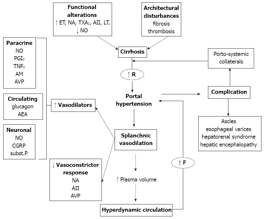
Figure 1 Physiopathology of portal hypertension.
In cirrhosis, the initiating factor leading to portal hypertension is an increase in intrahepatic vascular resistance (R), whereas the increase in portal blood flow (F) is a secondary phenomenon that maintains and worsens the increased portal pressure, giving rise to the hyperdynamic circulation syndrome. The different factors implicated in the distinct mechanisms of portal hypertension are shown. AII: angiotensin II; AEA: anandamide; AM: adrenomedullin; CGRP: calcitonine gene related peptide; CO: carbon monoxide; ET: endothelin; H2S: hydrogen sulfide; LT: leukotrienes; NE: norepinephrine; NO: nitric oxide; PGI2: prostacyclin; SP: substance P; TXA2: thromboxane A2.
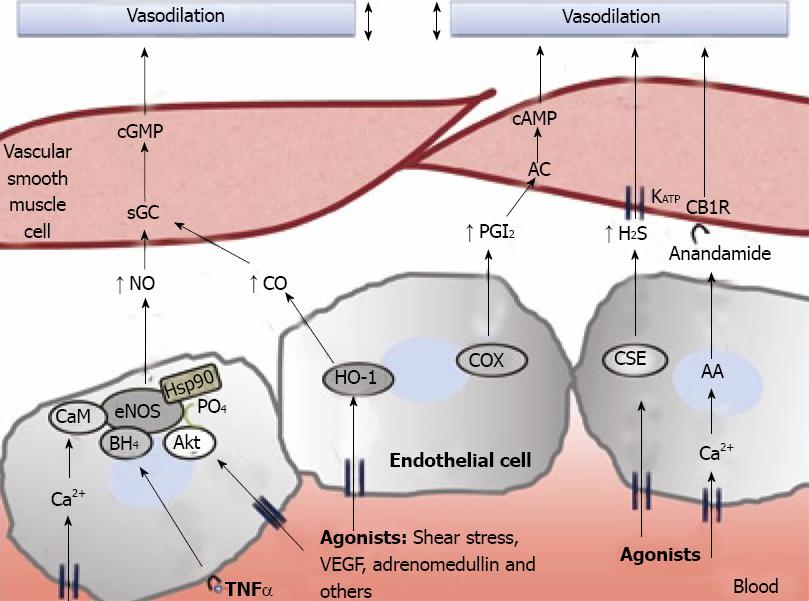
Figure 2 Molecular pathways associated to splanchnic vasodilation.
Vasoactive molecules involved in the regulation of vascular tone in the arteries of the splanchnic circulation. Nitric oxide (NO), carbon monoxide (CO), prostacyclin (PGl2) or hydrogen sulfide (H2S), generated through different pathways in endothelial cells, cause vasodilation in vascular smooth muscle cells by either activating soluble guanylate cyclase (sGC) to generate cyclic guanosine monophosphate (cGMP), by stimulating adenylate cyclase (AC) and generation of cyclic adenosine monophosphate (cAMP) or through the opening of KATP channels. Also, anandamide activates endothelial cannabinoid 1 receptors (CB1R) provoking vasodilation. AA: arachidonic acid; AC: adenylyl cyclase; Akt: protein kinase B; BH4: tetrahydrobiopterin; CaM: calmodulin; CSE: cystathionine-γ-lyase; COX: cyclooxygenase; eNOS: endothelial nitric oxide synthase; HSP90: heat shock protein 90; IP3: inositol triphosphate; TNFα: tumor necrosis factor α; VEGF: vascular endothelial growth factor.
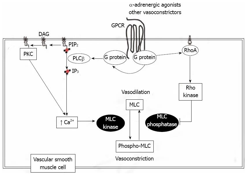
Figure 3 Vasodilation/contractile signaling in vascular smooth muscle cells.
The contractile state of vascular smooth muscle depends on the phosphorilation state of myosin light chains (MLCs). Under normal conditions, contractile agonists activate G protein couple receptors (GPCR). These receptors subsequently activate downstream effectors such as phospholipase C (PLC) and GTPase RhoA, leading to the increase of MLC phosphorilation via the activation of MLC kinase or the inhibition of MLC phosphatase. DAG: diacylglycerol; IP3: inositol triphosphate; PIP2: Phosphatidylinositol 4,5-bisphosphate; PKC: protein kinase C.
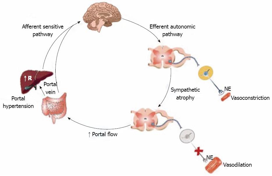
Figure 4 Hypothesis regarding the mechanisms and effects of the sympathetic post-ganglionic atrophy in splanchnic vasodilation.
The afferent stimulus of portal hypertension, originating from pressure increases in portal or mesenteric vessels or microvasculature, reaches the brain stem cardiovascular nuclei through the afferent nerves. From there, post-ganglionic sympathetic nerve regression are mediated by efferent sympathetic nerves, leading to neurotransmission inhibition and vasoconstriction impairment mediated by norepinephrine (NE).
- Citation: Martell M, Coll M, Ezkurdia N, Raurell I, Genescà J. Physiopathology of splanchnic vasodilation in portal hypertension. World J Hepatol 2010; 2(6): 208-220
- URL: https://www.wjgnet.com/1948-5182/full/v2/i6/208.htm
- DOI: https://dx.doi.org/10.4254/wjh.v2.i6.208


