Copyright
©2010 Baishideng.
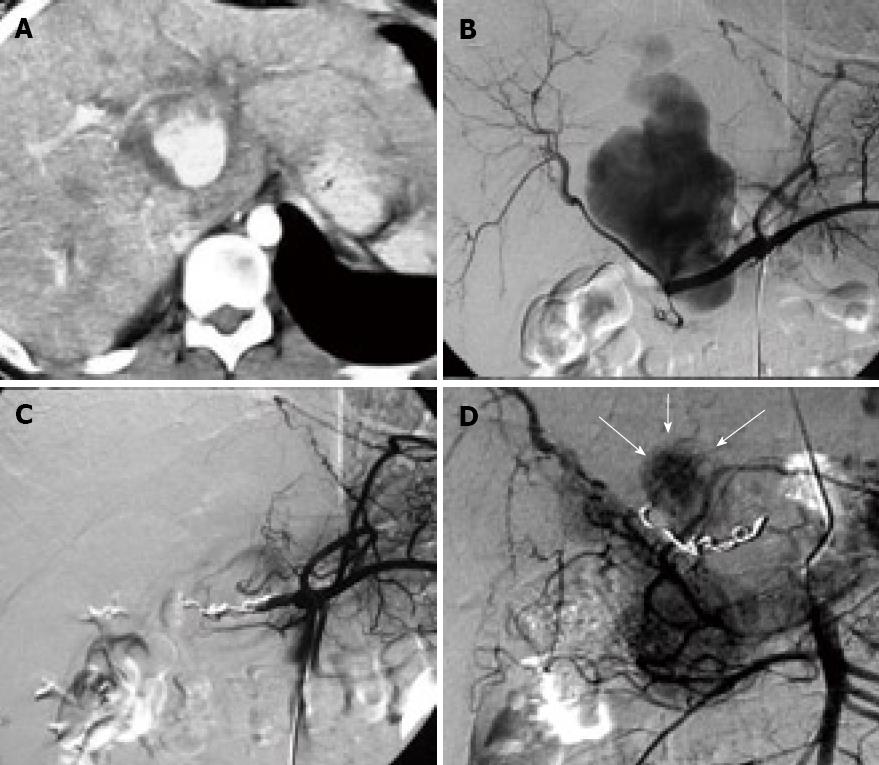
Figure 1 CT and angiography images.
A: contrast enhanced CT revealing a collection filled of contrast at the hepatic hilium compatible with pseudoaneurysm and dilated intrahepatic bile duct; B: Selective arteriography of the celiac trunk showing the pseudoaneurysm arising from the common hepatic artery; C: Angiography of the common hepatic artery after embolization. No fill of the pseudoaneurysm is visible from this artery; D: Selective superior mesenteric artery arteriography three days after embolization. The pseudoaneurysm (white arrows) is partly thrombosed with persistence of filling from thin branches. The superselective catheterization of these vessel wasn´t possible due its tortuosity and narrow caliber.
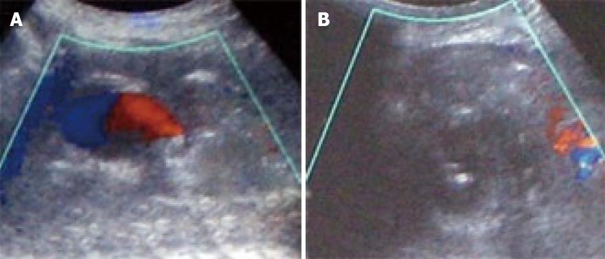
Figure 2 Ultrasound guided thrombin injection.
A: Color doppler ultrasound. A small cavity persists with flow in the pseudoaneurysm; B: Needle inside the pseudoaneurysm immediately after the injection of thrombin showing the absence of flow (thrombosis).
Figure 3 CT scan 12 mo after treatment.
Complete thrombosis of the pseudoaneurysm and no dilation of the biliary tract are observed.
- Citation: Francisco LE, Asunción LC, Antonio CA, Ricardo RC, Manuel RP, Caridad MH. Post-traumatic hepatic artery pseudoaneurysm treated with endovascular embolization and thrombin injection. World J Hepatol 2010; 2(2): 87-90
- URL: https://www.wjgnet.com/1948-5182/full/v2/i2/87.htm
- DOI: https://dx.doi.org/10.4254/wjh.v2.i2.87


