Copyright
©2010 Baishideng.
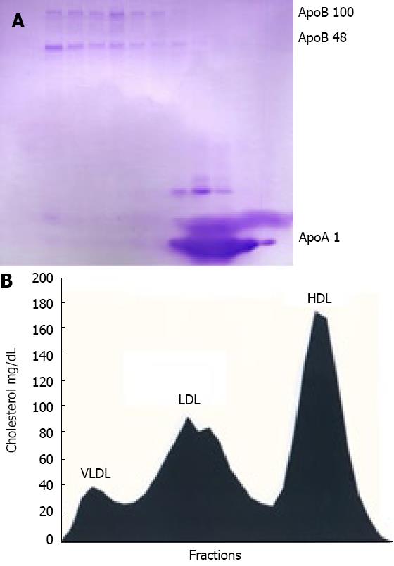
Figure 1 FPLC fractionation of mouse plasma.
Mice kept on high-fat diet were bled and pooled plasma was adjusted with KBr to d = 1.215 g/mL and ultracentrifuged. Resulting lipoprotein fraction with density d < 1.215 g/mL was subjected to FPLC gel filtration and cholesterol content was measured in each fraction. Three of each consecutive fraction were pooled, delipidated, heat-denatured and subjected to SDS PAGE gel electrophoresis in gradient gel as described in “Material and Methods”. A: Coomassie blue-stained gel with ApoB-100, ApoB-48 and ApoAI indicated by arrow; B: Cholesterol levels in FPLC fractions aligned to apolipoprotein profile of mouse plasma.
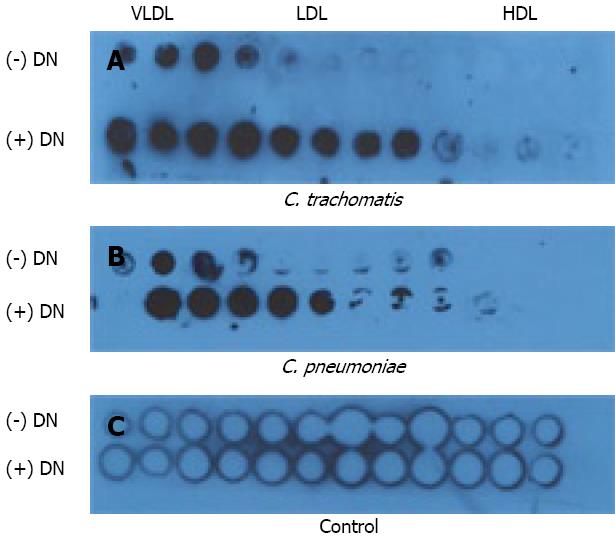
Figure 2 Immuno-dot blot binding assay.
Heat-denatured (+ DN) or non-heat-denatured (- DN) FPLCS fractions of mouse plasma lipoproteins were applied to nitrocellulose membrane and incubated with elementary bodies of C. trachomatis (Panel A) or C. pneumoniae (Panel B) and blotted with chlamydial LPS-specific antibodies as described in “Material and Methods”. Control membrane (Panel C) treated similarly except addition of chlamydial suspension.
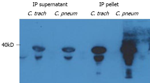
Figure 3 Immunoprecipitation analysis.
Soluble membrane extracts of C. trachomatis and C. pneumoniae preincubated with native Apo-B containing lipoproteins were immunoprecipitated with Apo-B specific polyclonal antibody and subjected to SDS PAGE. Immunoprecipitated (IP) supernatant and pellet were obtained as described in Materials and Methods. Membranes were immunoblotted with MOMP-specific monoclonal antibodies.
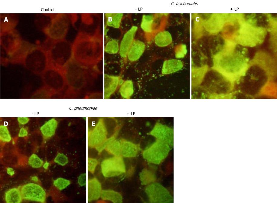
Figure 4 HepG2 cells.
Direct immunofluorescence with FITC-conjugated antibody against chlamydial LPS. HepG2 cells were infected with inoculants containing: A: No chlamydial particles, B: C. trachomatis alone; C: C. trachomatis preincubated with native Apo-B containing lipoproteins; D: C. pneumoniae alone; E: C. pneumoniae preincubated with native Apo-B containing lipoproteins. Immunofluorescence protocol is detailed in Materials and Methods. Original magnification × 40.
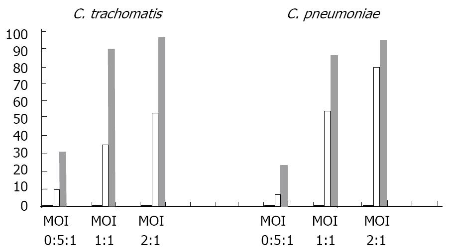
Figure 5 Flow Cytometry Analysis of HepG2 cells.
Infectivity of C. pneumoniae and C. trachomatis preincubated with native ApoB-containing lipoproteins. HepG2 cells were infected with C. pneumoniae (left side of diagram) or C. trachomatis (right side of diagram) with or without preincubation of bacteria with native ApoB-containing lipoproteins as described in Material Methods. Three different levels of infection multiplicity were studied for each pathogen (MOI 0.5:1; 1:1; 2:1). Percentage of the infected cells shown on value Y axis was determined by flow cytometry (see Materials and Methods) for control (uninfected) HepG2 suspensions (hardly seen black columns), HepG2 suspensions infected with chlamydia bacteria alone (white columns) and HepG2 suspensions infected with chlamydial bacteria preincubated with native ApoB-containing lipoproteins (gray columns).
- Citation: Bashmakov YK, Zigangirova NA, Gintzburg AL, Bortsov PA, Petyaev IM. ApoB-containing lipoproteins promote infectivity of chlamydial species in human hepatoma cell line. World J Hepatol 2010; 2(2): 74-80
- URL: https://www.wjgnet.com/1948-5182/full/v2/i2/74.htm
- DOI: https://dx.doi.org/10.4254/wjh.v2.i2.74


