
















Copyright: ©Author(s) 2026.
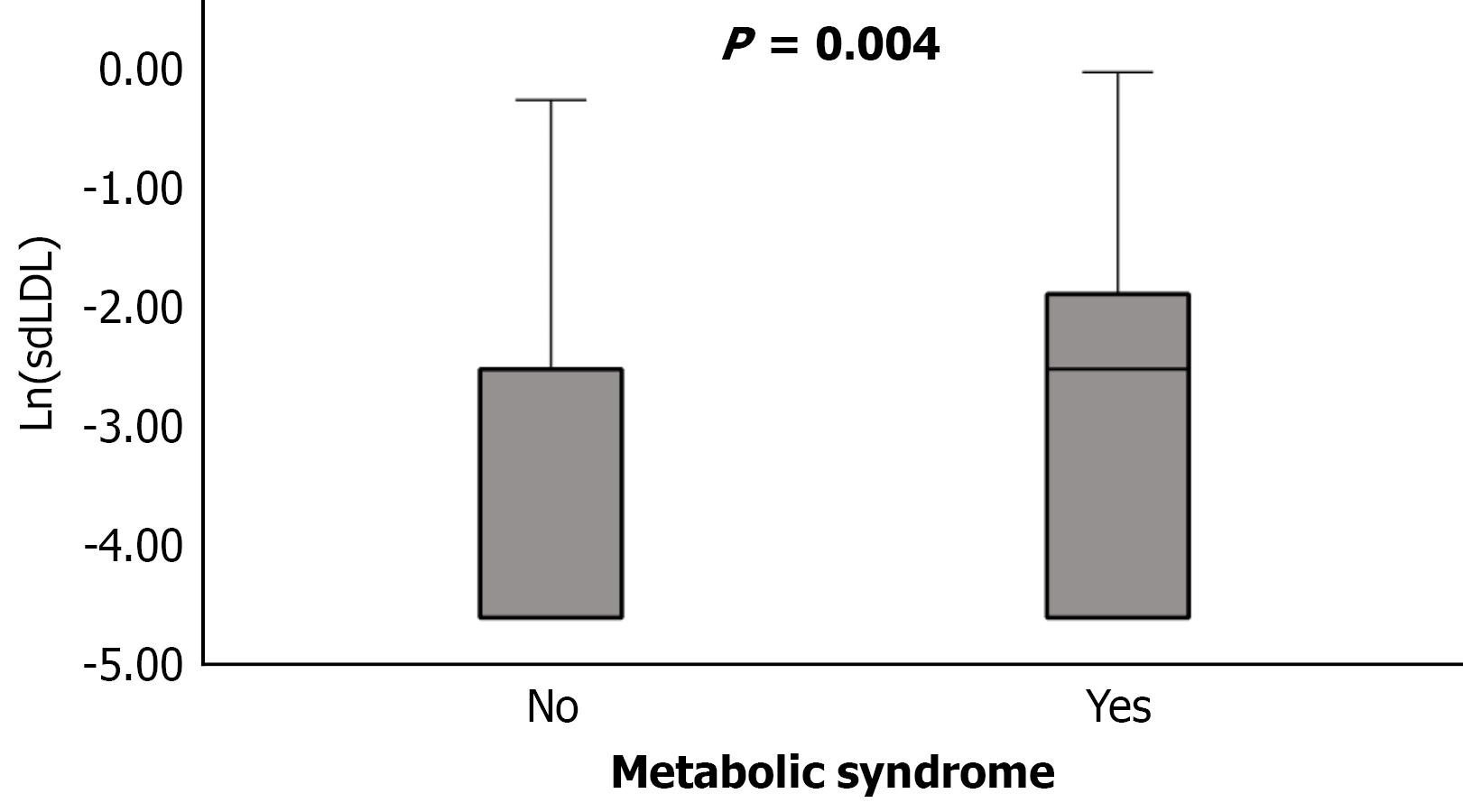
Figure 1 Distribution of ln-transformed small dense low-density lipoprotein levels according to metabolic syndrome status.
Ln (small dense low-density lipoprotein) levels were significantly higher in patients with metabolic syndrome than in those without metabolic syndrome (P = 0.004, Mann-Whitney U test). Boxplots represent median values, interquartile ranges, and whiskers. sdLDL: Small dense low-density lipoprotein.
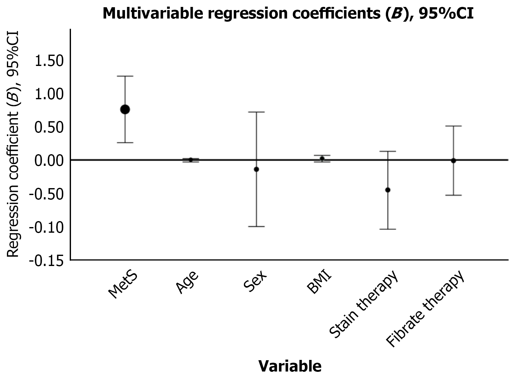
Figure 2 Multivariable linear regression analysis of factors associated with ln-transformed small dense low-density lipoprotein levels.
Error bars represent 95% confidence interval. Metabolic syndrome remained independently associated with higher ln (small dense low-density lipoprotein) levels after adjustment for age, sex, body mass index, and lipid-lowering therapy. BMI: Body mass index; CI: Confidence interval.
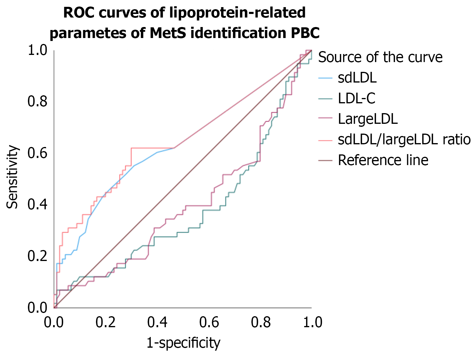
Figure 3 Receiver operating characteristic analysis of lipoprotein-related parameters for identification of metabolic syndrome.
Receiver operating characteristic curves are shown for small dense low-density lipoprotein (sdLDL), low-density lipoprotein (LDL)-C, large LDL, and sdLDL/LargeLDL ratio. The sdLDL/LargeLDL ratio demonstrated the highest discriminatory performance, whereas LDL-C and large LDL showed inverse discriminatory patterns. Curves for LDL-C and large LDL fall below the diagonal reference line, indicating that lower values of these parameters were associated with metabolic syndrome. sdLDL: Small dense low-density lipoprotein; ROC: Receiver operating characteristic; MetS: Metabolic syndrome; LDL: Low-density lipoprotein; PBC: Primary biliary cholangitis.
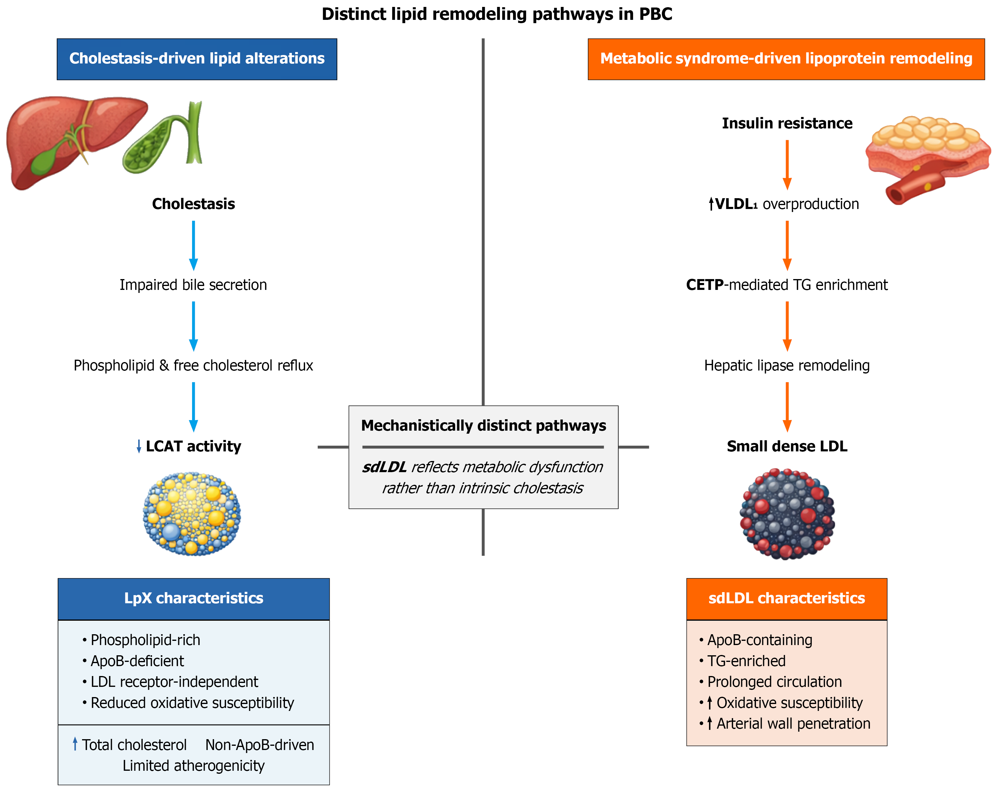
Figure 4 Mechanistically distinct lipid remodeling pathways in primary biliary cholangitis.
Left panel: Cholestasis impairs bile acid secretion and disrupts biliary phospholipid transport, leading to reflux of phospholipids and free cholesterol into the circulation and reduced lecithin-cholesterol acyltransferase activity. These alterations promote the formation of lipoprotein X, a phospholipid-rich, apolipoprotein B (ApoB)-deficient particle that contributes to hyper
- Citation: Koky T, Drazilova S, Janicko M, Harhovsky M, Hubkova B, Tothova I, Rabajdova M, Marekova M, Jarcuska P. Small dense low-density lipoprotein in primary biliary cholangitis: Independent association with metabolic syndrome. World J Hepatol 2026; 18(8): 124436
- URL: https://www.wjgnet.com/1948-5182/full/v18/i8/124436.htm
- DOI: https://dx.doi.org/10.4254/wjh.124436