
















Copyright: ©Author(s) 2026.
World J Hepatol. Oct 27, 2026; 18(10): 125241
Published online Oct 27, 2026. doi: 10.4254/wjh.125241
Published online Oct 27, 2026. doi: 10.4254/wjh.125241
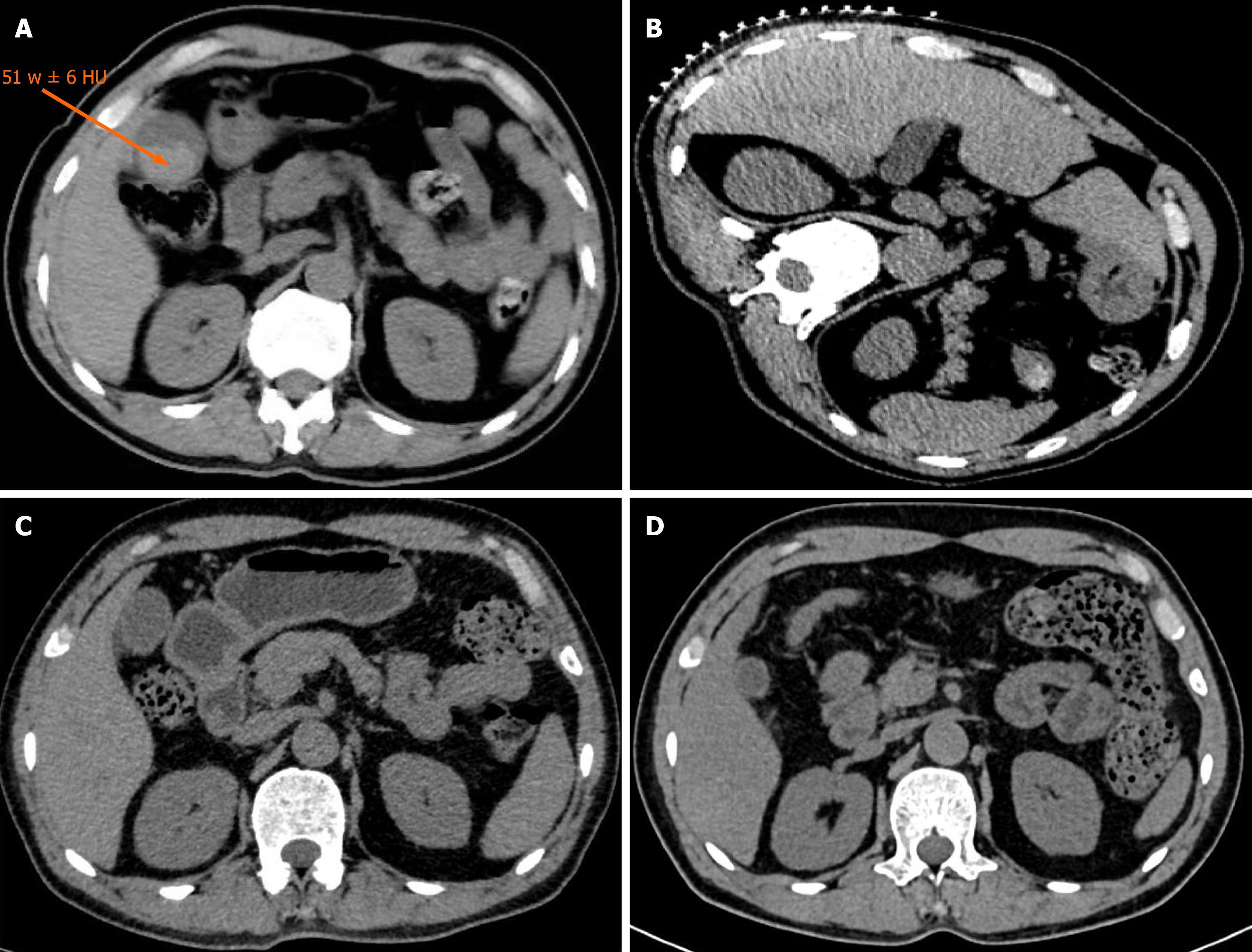
Figure 1 Dynamic computed tomography progression of acute hemobilia following computed tomography-guided percutaneous liver biopsy.
A: Acute phase of complication (immediately post-withdrawal). An axial unenhanced computed tomography (CT) scan demonstrates significant gallbladder distension compared to the preoperative baseline, filled with diffuse, homogeneous intraluminal hyperdensity (mean CT attenuation: 51 ± 6 Hounsfield units; arrow), consistent with acute intraluminal hemorrhage; B: Preoperative baseline comparison. The baseline CT image obtained prior to the biopsy shows a physiologically collapsed gallbladder with no evidence of intraluminal abnormalities or prior biliary disease; C: Stable observation phase (5 days post-procedure). A repeat CT scan reveals stabilized gallbladder dimensions and persistent intraluminal hyperdensity without further expansion, indicating a contained and non-progressive hemobilia; D: Follow-up absorption phase (1 month post-procedure). A follow-up CT scan demonstrates restoration of normal gallbladder morphology with complete absorption of the intraluminal hematoma.
- Citation: Xu YC, Halimulati T, Yu XF, Guo XX, Zhao DD, Bi JW, Wang Q. Acute hemobilia following computed tomography-guided percutaneous liver biopsy: A case report. World J Hepatol 2026; 18(10): 125241
- URL: https://www.wjgnet.com/1948-5182/full/v18/i10/125241.htm
- DOI: https://dx.doi.org/10.4254/wjh.125241