Copyright
©The Author(s) 2024.
World J Hepatol. Jun 27, 2024; 16(6): 966-972
Published online Jun 27, 2024. doi: 10.4254/wjh.v16.i6.966
Published online Jun 27, 2024. doi: 10.4254/wjh.v16.i6.966
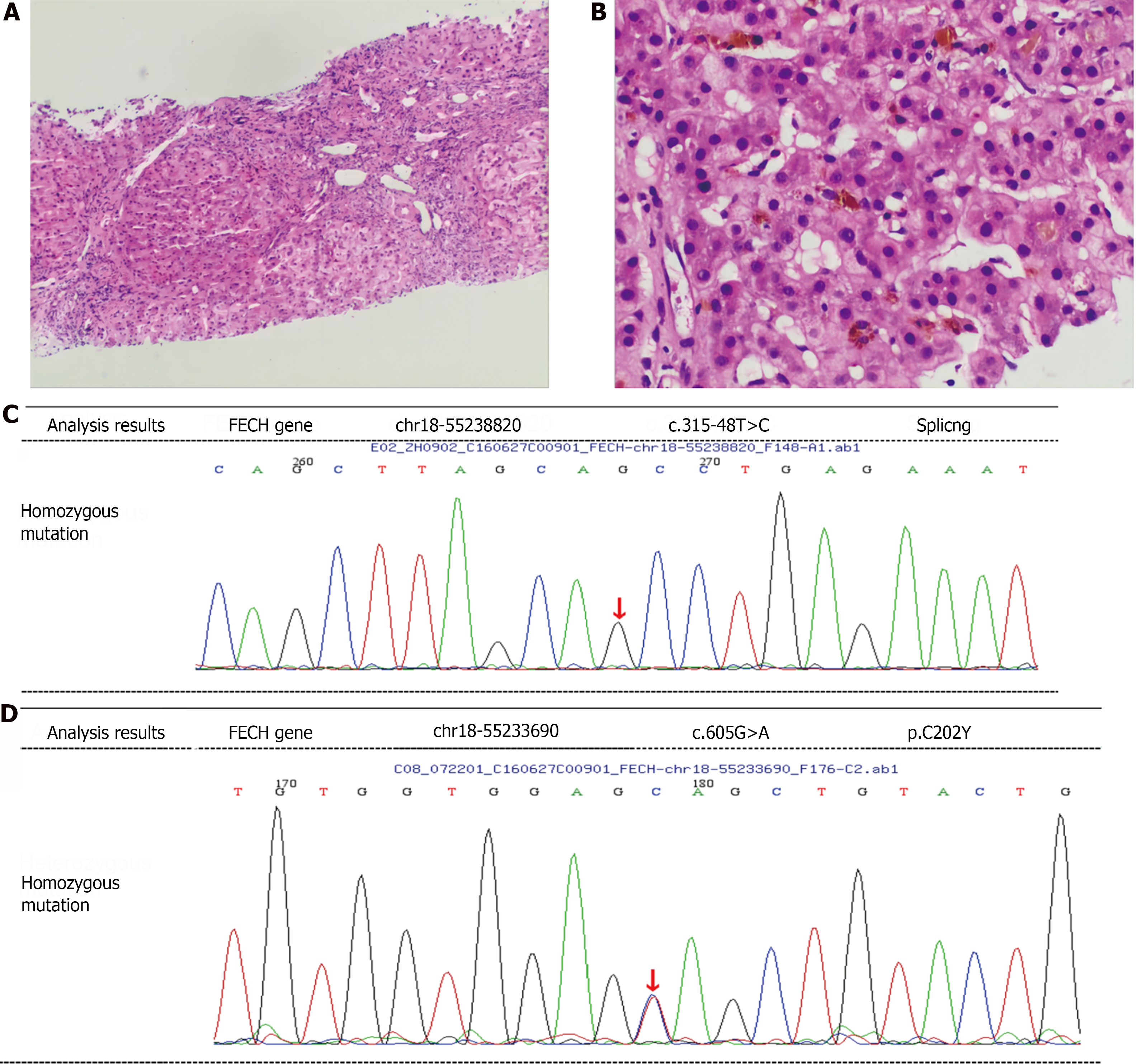
Figure 1 Hepatic pathology and genetic screening.
A and B: Liver histopathology demonstrates brown-yellow granule deposits in hepatocytes, capillary ducts, and Kupffer cells within the hepatic sinusoids. Enlarged portal area with increased lymphocyte and neutrophil infiltration, fibrous tissue hyperplasia, and hyperplasia of small bile ducts consistent with G3S3 chronic inflammatory liver injury; C and D: Genetic analysis reveals ferrochelatase gene mutations: homozygous intron c.315-48T>C mutation and heterozygous p.C202Y mutation. FECH: Ferrochelatase.
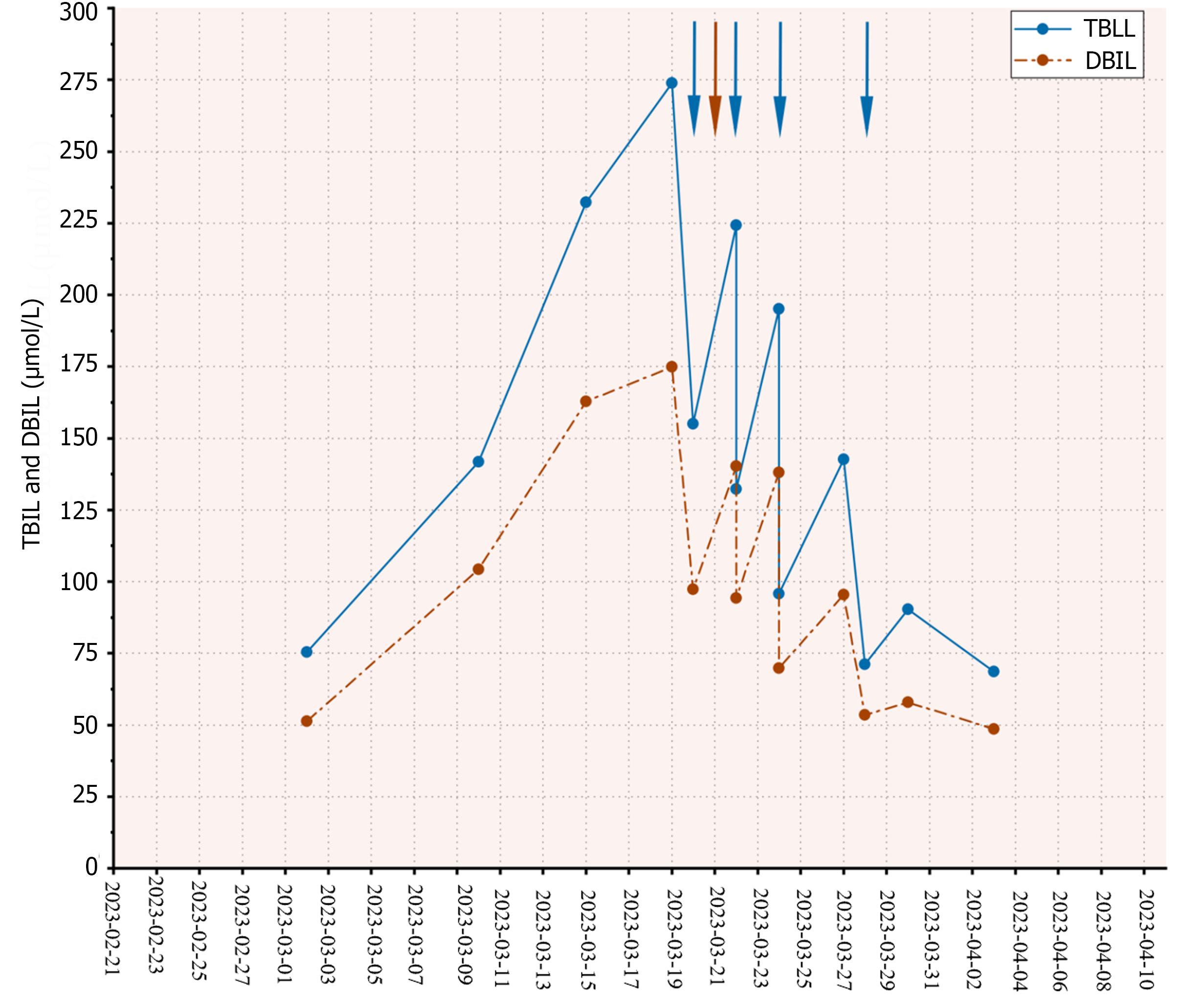
Figure 2 Dynamic changes in total bilirubin and direct bilirubin levels in the patient.
Plasma exchange treatments were conducted on March 20, 22, 24, and 28, 2023 (involving the replacement of 1800-2000 mL of fresh frozen plasma during each session), and a red blood cell exchange treatment was performed on March 21, 2023 (approximately 6 units of red blood cells were transfused). Blue arrows represent plasma exchange procedures while brown arrow denotes red blood cell exchange treatments. TBIL: Total bilirubin; DBIL: Direct bilirubin.
- Citation: Zeng T, Chen SR, Liu HQ, Chong YT, Li XH. Successful treatment of severe hepatic impairment in erythropoietic protoporphyria: A case report and review of literature. World J Hepatol 2024; 16(6): 966-972
- URL: https://www.wjgnet.com/1948-5182/full/v16/i6/966.htm
- DOI: https://dx.doi.org/10.4254/wjh.v16.i6.966


