Copyright
©The Author(s) 2023.
World J Hepatol. Feb 27, 2023; 15(2): 201-207
Published online Feb 27, 2023. doi: 10.4254/wjh.v15.i2.201
Published online Feb 27, 2023. doi: 10.4254/wjh.v15.i2.201
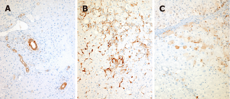
Figure 1 Immunohistochemical staining of normal, non-alcoholic steatohepatitis, and non-alcoholic steatohepatitis cirrhosis liver tissue with a galectin-3 antibody.
A: Normal liver: In the normal liver, immunoreactive galectin-3 is present in epithelial cells of all large and small bile ducts and in periportal ductules; B: Non-alcoholic steatohepatitis (NASH) liver: In livers with inflammatory disease, staining appears in activated macrophages both in the parenchyma and in the portal tracts as well as in lymphoid germinal centers when present in chronic hepatitis. Notably there is no galectin-3 positivity in hepatocytes in pre-cirrhotic inflammatory disease, even in the presence of ballooning degeneration, Mallory-Denk bodies or features of apoptosis; C: NASH cirrhosis: Galectin-3 appears in hepatocytes and can be focal or widespread with both cytoplasmic and nuclear activity. A-C: Citation: Goodman Z, Lowe E, Boudes P. Hepatic Expression of Galectin-3, a Pro-fibrotic and Pro-inflammatory Marker: An Immunohistochemical Survey. Proceedings of the American Association for the Study of Liver Diseases meetings; 2022 Nov; Washington, US. Copyright© The Authors 2020. The authors have obtained the permission for figure using from the authors (Supplementary material).
- Citation: Kram M. Galectin-3 inhibition as a potential therapeutic target in non-alcoholic steatohepatitis liver fibrosis. World J Hepatol 2023; 15(2): 201-207
- URL: https://www.wjgnet.com/1948-5182/full/v15/i2/201.htm
- DOI: https://dx.doi.org/10.4254/wjh.v15.i2.201


