Copyright
©The Author(s) 2023.
World J Hepatol. Nov 27, 2023; 15(11): 1196-1209
Published online Nov 27, 2023. doi: 10.4254/wjh.v15.i11.1196
Published online Nov 27, 2023. doi: 10.4254/wjh.v15.i11.1196
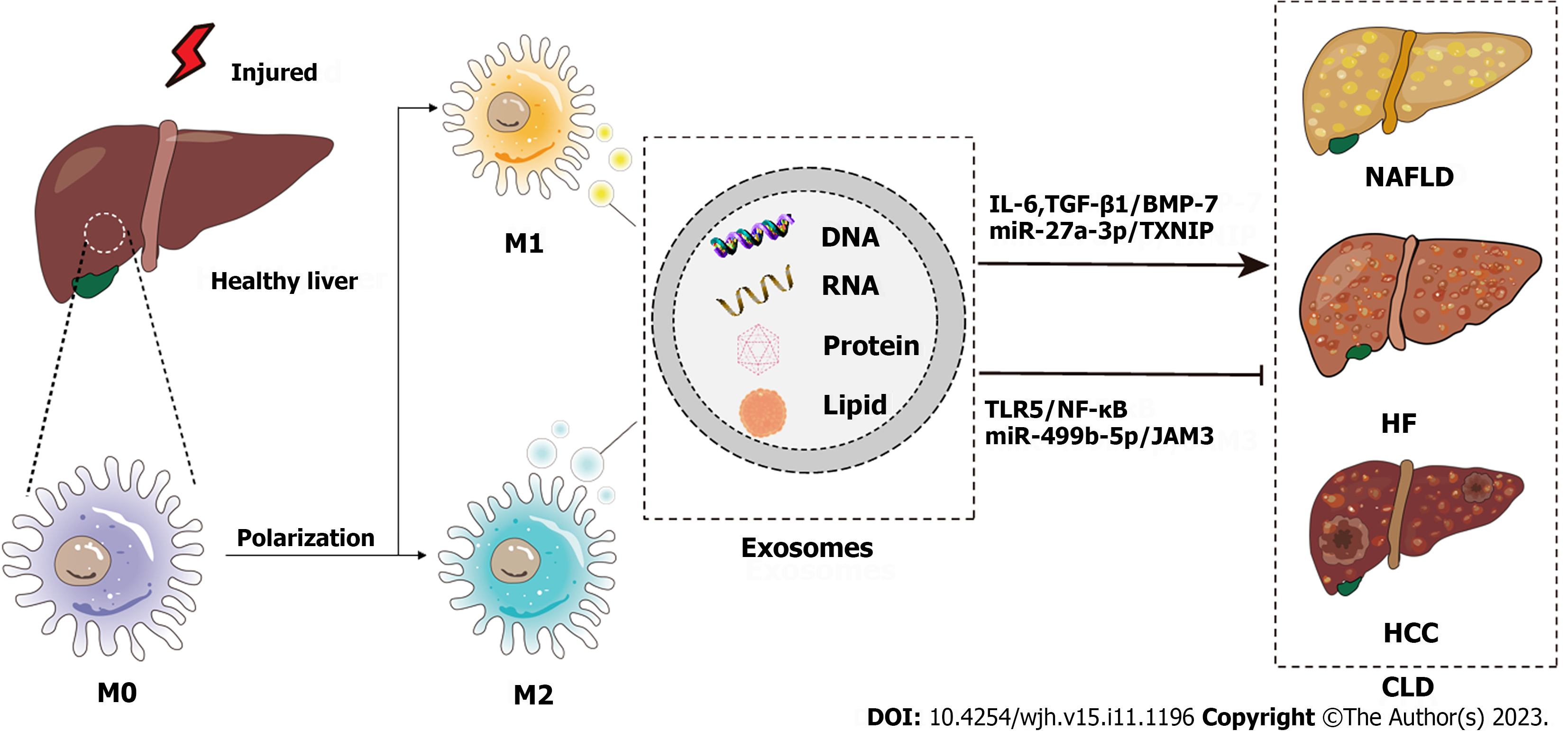
Figure 1 Schematic diagram of the pathogenesis of chronic liver disease from the perspective of macrophage-derived exosomes.
Injured livers activate macrophages to secrete exosomes that encapsulate RNAs, DNAs, lipids, proteins, etc., which influence the development of chronic liver disease through various signaling pathways. CLD: Chronic liver disease; NAFLD: Nonalcoholic fatty liver disease; HF: Hepatic fibrosis; HCC: Hepatocellular carcinoma; IL-6: Interleukin-6; TGF: Transforming growth factor; BMP: Bone morphogenetic protein; TXNIP: Thioredoxin-interacting protein; TLR: Toll-like receptor.
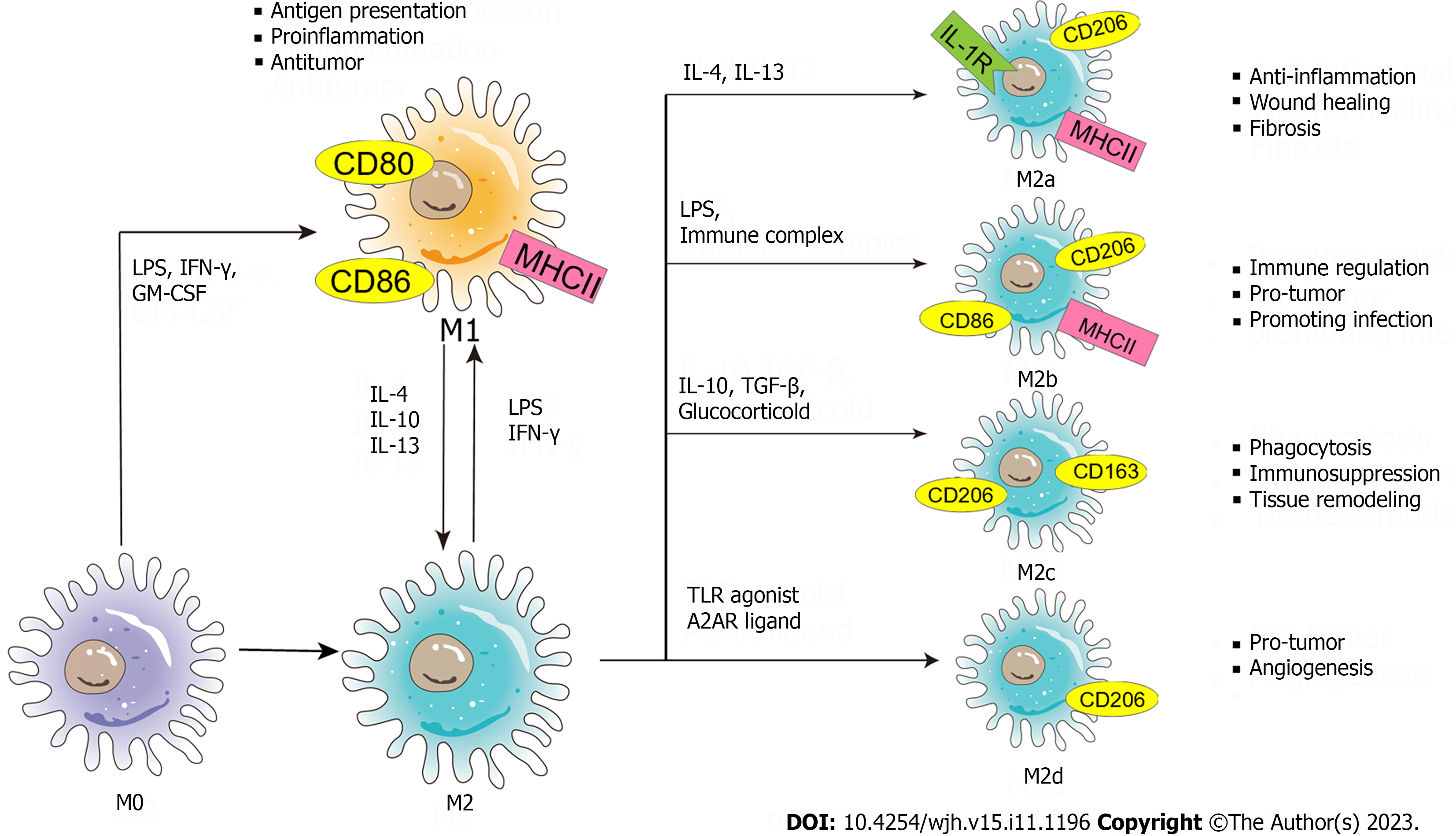
Figure 2 Schematic diagram of the phenotypes and functions of macrophage polarization.
The nature macrophage can be activated by a variety of influencing factors (such as lipopolysaccharide, interferon-γ, granulocyte-macrophage colony-stimulating factor, etc.) and polarized into two phenotypes - classically activated macrophages and alternatively activated macrophages. Exosomes carry stimulatory factors to activate macrophages. Macrophages themselves secrete exosomes to form a signal transmission network between macrophages and other cells. (M0: M0 macrophage; M1: M1 macrophage; M2: M2 macrophage; M2a: M2a macrophage; M2b: M2b macrophage; M2c: M2c macrophage; M2d: M2d macrophage; LPS: Lipopolysaccharide; IFN-γ: Interferon-γ; GM-CSF: Granulocyte-macrophage colony-stimulating factor; IL: Interleukin; TLR: Toll-like receptor; A2AR: A2A receptor; MHC: Major histocompatibility complex; TGF: Transforming growth factor.
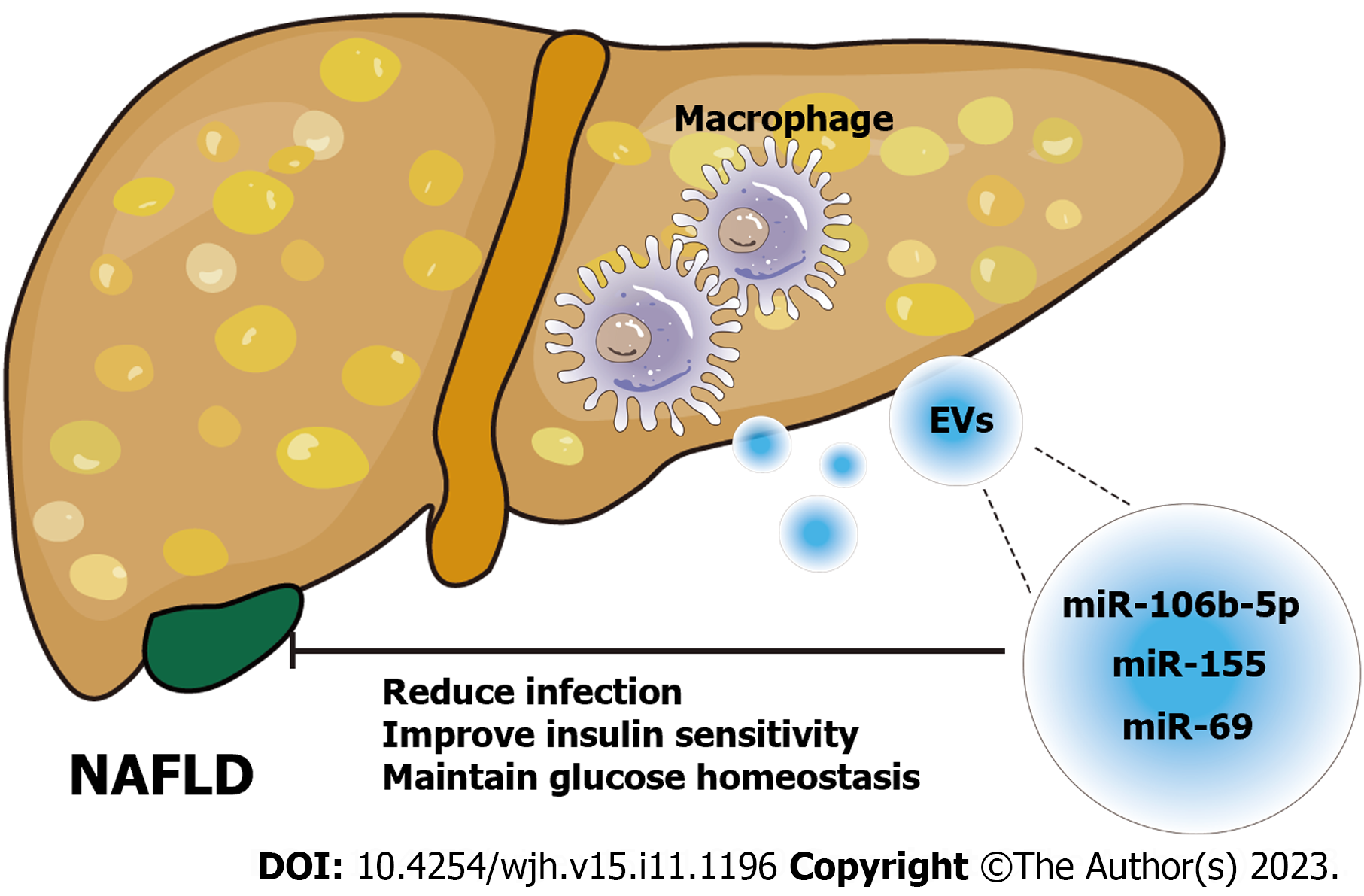
Figure 3 Schematic diagram of the role of macrophage-derived exosomes in nonalcoholic fatty liver disease.
Exosomes derived from macrophages carry different microRNAs (miR-106b-5p[63], miR-155[64], miR-69[65]) to act on nonalcoholic fatty liver disease cells and alleviate the disease progression by regulating liver homeostasis. NAFLD: Nonalcoholic fatty liver disease; EVs: Extracellular vesicles; miRNA: MicroRNA.
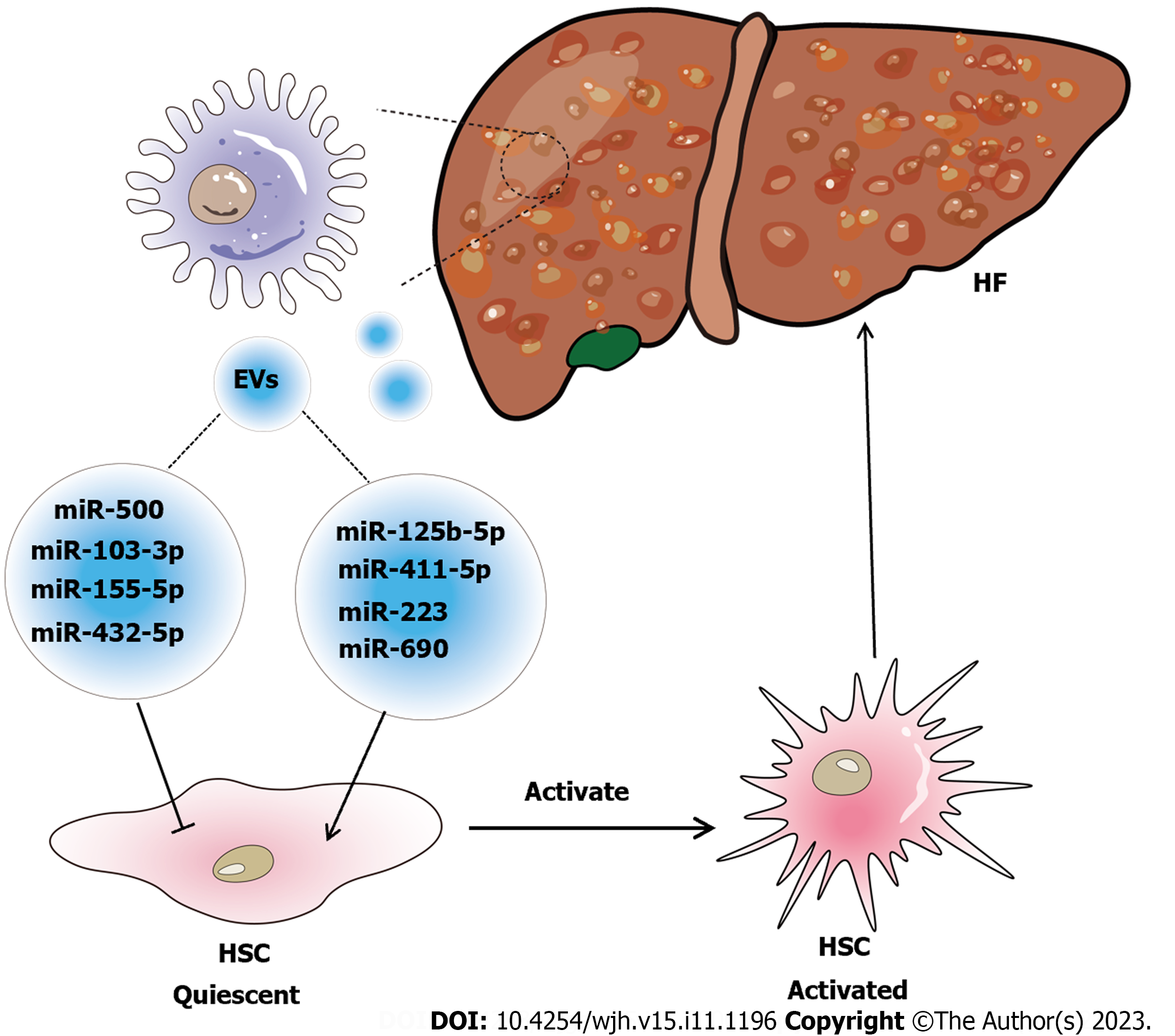
Figure 4 Schematic diagram of the relationship between macrophage-derived exosomes and hepatic fibrosis.
Exosome-mediated communication between macrophages and hepatic stellate cell (HSC) influences the disease progression of hepatic fibrosis (HF). Special substances carried in exosomes, such as microRNAs (miR-103-3p[24]; miR-500[70]; miR-155-5p[72]; miR-432-5p[73]; miR-125b-5p[74]; miR-411-5p[75]; miR-690[65]; miR-223[23], are signaling factors that induce the activation of HSC to form cirrhosis or inhibit the activation of HSC to alleviate HF. EVs: Extracellular vesicles; HSC: Hepatic stellate cell; HF: Hepatic fibrosis.
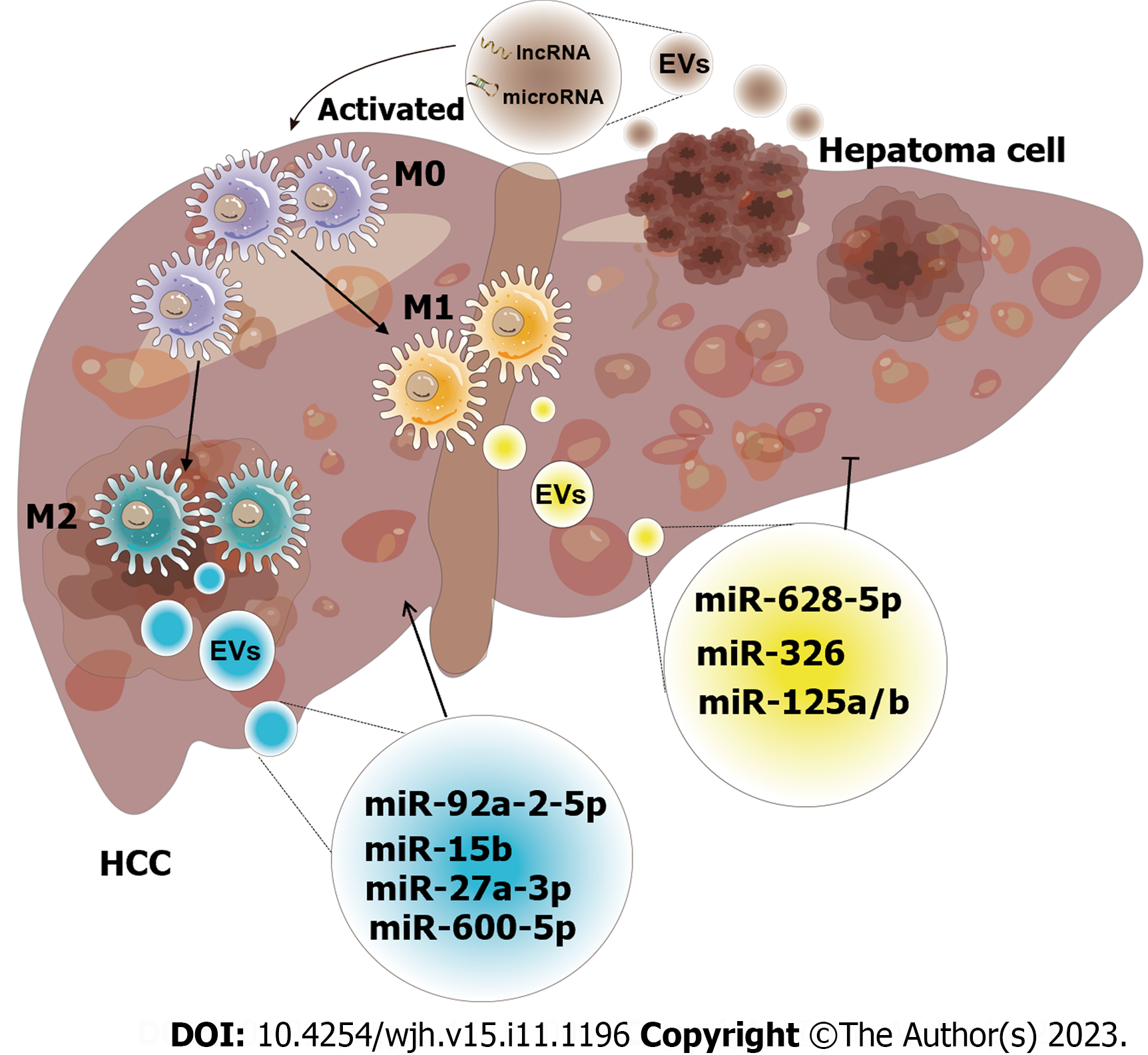
Figure 5 Schematic diagram of the effect of exosomes derived from macrophages on hepatocellular carcinoma.
The crosstalk of exosomes between macrophages and hepatocellular carcinoma (HCC) affects tumor progression. Both exosomes released by macrophages and HCC have unique intracellular components, including a variety of mRNAs, microRNAs, long non-coding RNAs, lipids, etc. These intracellular components are utilized as communicators to induce pathways that result in increased or inhibited cell proliferation, invasion, and other hallmarks of malignancy (M0: M0 macrophage; M1: M1 macrophage; M2: M2 macrophage; miR-92a-2-5p[82]; miR-15b[83]; miR-27a-3p[85]; miR-660-5p[86]; miR-628-5p[87]; miR-326[88]; miR-125a/b[90]). EVs: Extracellular vesicles; HCC: Hepatocellular carcinoma; miRNA: MicroRNA; lncRNA: Long non-coding RNA.
- Citation: Xiang SY, Deng KL, Yang DX, Yang P, Zhou YP. Function of macrophage-derived exosomes in chronic liver disease: From pathogenesis to treatment. World J Hepatol 2023; 15(11): 1196-1209
- URL: https://www.wjgnet.com/1948-5182/full/v15/i11/1196.htm
- DOI: https://dx.doi.org/10.4254/wjh.v15.i11.1196


