Copyright
©The Author(s) 2022.
World J Hepatol. Jul 27, 2022; 14(7): 1512-1519
Published online Jul 27, 2022. doi: 10.4254/wjh.v14.i7.1512
Published online Jul 27, 2022. doi: 10.4254/wjh.v14.i7.1512
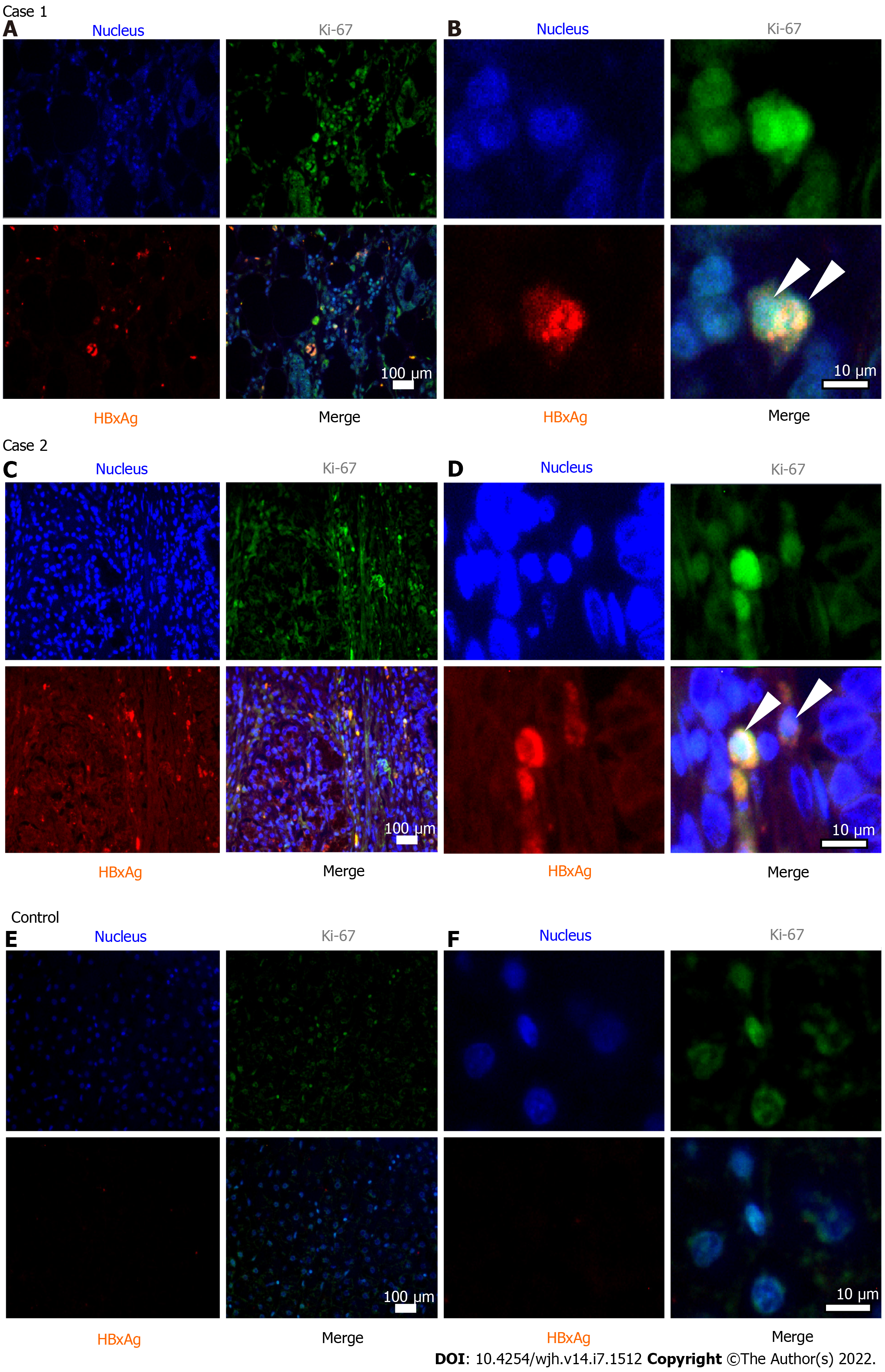
Figure 1 Immunohistochemistry of resected pancreatic tissues.
Case 1 and case 2 are subjects with pancreatic ductal adenocarcinoma and positive markers of hepatitis B virus (HBV) infection. Control refers to pancreatic tissue of a patient with pancreatic cancer, negative for markers of current and previous HBV infection (control case is not described). Samples were stained for Ki-67 protein (green fluorescence) and HBV regulatory X protein (HBx) (red fluorescence). Cell nuclei were counterstained with Hoechst33342 dye (blue). A, C and E: Images at magnification 10 ×; B, D and F: Images at magnification 100 ×. Arrows indicate HBx/Ki-67 co-stained cells. Median Ki-67 index (%): Subject 1 - 77, Subject 2 - 68, Сontrol - 55.
- Citation: Batskikh S, Morozov S, Kostyushev D. Hepatitis B virus markers in hepatitis B surface antigen negative patients with pancreatic cancer: Two case reports . World J Hepatol 2022; 14(7): 1512-1519
- URL: https://www.wjgnet.com/1948-5182/full/v14/i7/1512.htm
- DOI: https://dx.doi.org/10.4254/wjh.v14.i7.1512


