Copyright
©The Author(s) 2022.
World J Hepatol. Feb 27, 2022; 14(2): 338-353
Published online Feb 27, 2022. doi: 10.4254/wjh.v14.i2.338
Published online Feb 27, 2022. doi: 10.4254/wjh.v14.i2.338
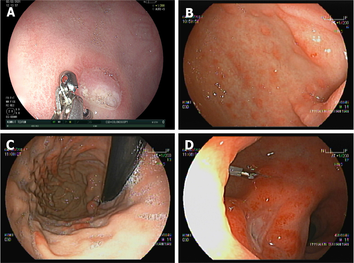
Figure 1 Cytomegalovirus tissue infection of the stomach and duodenum in a 13-mo-old boy and a 14-year-old boy with D+/R− serostatus at transplant.
Neither patient received antiviral prophylaxis. A and B: The 13-mo-old boy with D+/R− serostatus at transplant presented with severe anaemia at 3 mo; C and D: The 14-year-old boy presented with haematemesis at 2 mo after liver transplantation.
Figure 2 Biopsies showing chronic active gastritis.
A: Cytomegalovirus inclusion bodies are seen within mucous cells. The gastric biopsy is characterized by enlarged cells with basophilic nuclear and cytoplasmic inclusions; B: Liver biopsy shows a neutrophilic microabscess surrounding a hepatocyte with granular basophilic cytoplasmic cytomegalovirus inclusions; C: Positive cytomegalovirus immunohistochemistry in liver tissue.
- Citation: Onpoaree N, Sanpavat A, Sintusek P. Cytomegalovirus infection in liver-transplanted children. World J Hepatol 2022; 14(2): 338-353
- URL: https://www.wjgnet.com/1948-5182/full/v14/i2/338.htm
- DOI: https://dx.doi.org/10.4254/wjh.v14.i2.338


