©The Author(s) 2021.
World J Hepatol. Jan 27, 2021; 13(1): 66-79
Published online Jan 27, 2021. doi: 10.4254/wjh.v13.i1.66
Published online Jan 27, 2021. doi: 10.4254/wjh.v13.i1.66
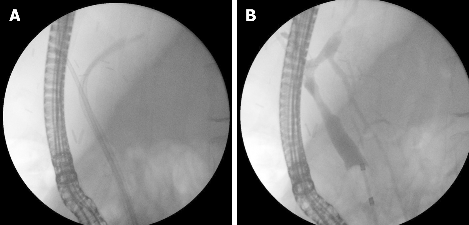
Figure 1 Endoscopic treatment of anastomotic stricture after living donor liver transplantation.
A: Two plastic stents; and B: Occlusive cholangiogram after treatment.
Figure 2 Anastomotic stricture.
A: Cholangiogram; B: Balloon dilation; and C: Multiple stent treatment.
Figure 3 Anastomotic stricture after living donor liver transplantation (right lobe).
A: Guidewire insertion; B: Balloon dilation; C: Second guidewire insertion; and D: Stent placement (7Fr + 5Fr).
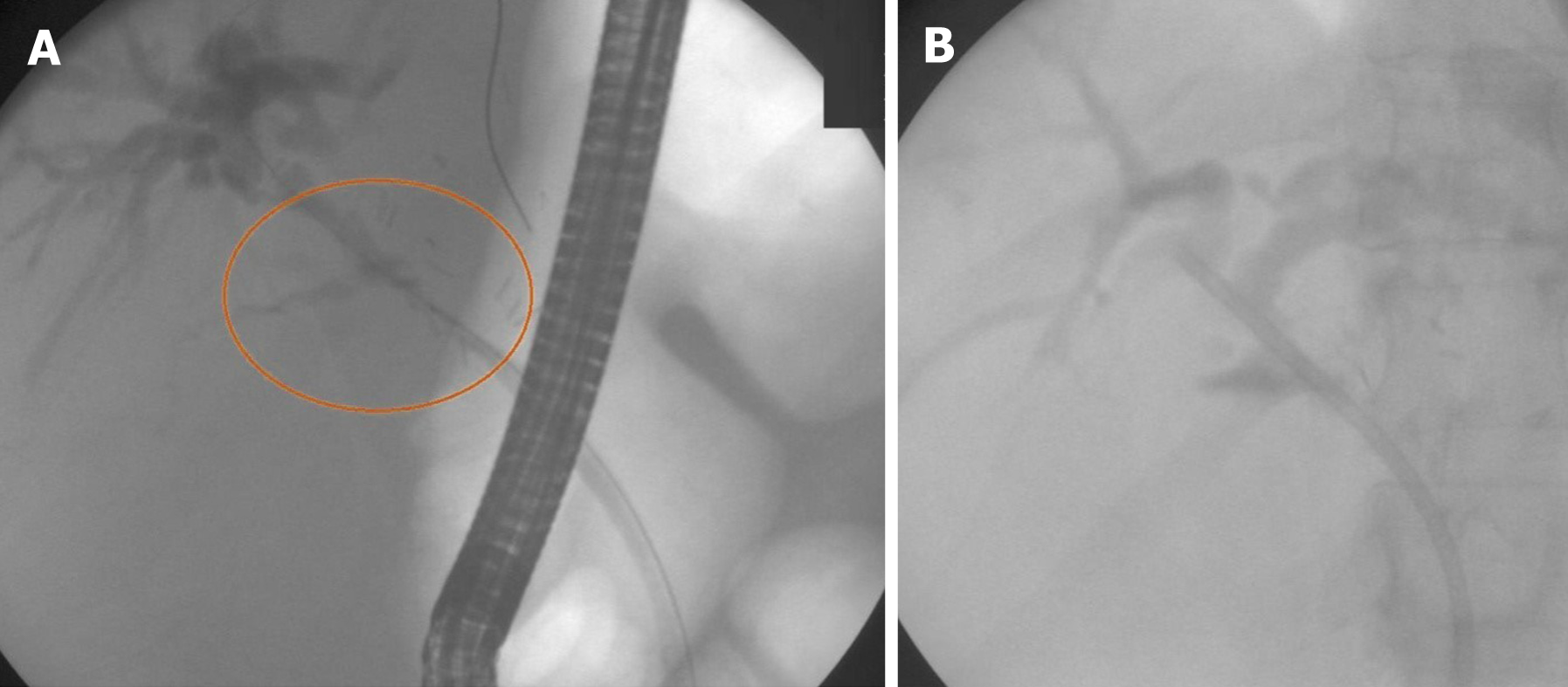
Figure 4 Complex anastomotic stricture.
A: Impossible insertion of guidewire through a stricture; B: Guidewire insertion under direct visual control; and C: Guidewire inserted above anastomosis.
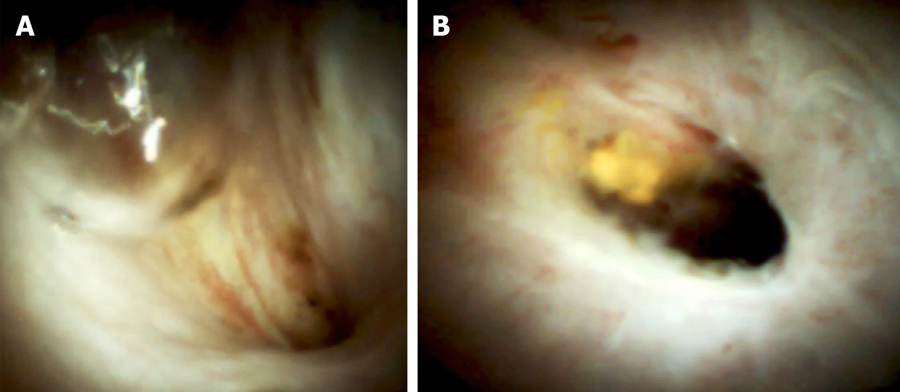
Figure 5 Digital cholangioscopy image of an anastomotic stricture.
Figure 6 Anastomotic leak.
A: Guidewire insertion; and B: Stent placement (10Fr).
Figure 7 Multiple intrahepatic stones above anastomotic stricture.
A: Fluoroscopic image; B: Digital cholangioscopic image; C: Electrohydraulic lithotripsy performance; and D: Fluoroscopic image after treatment.
Figure 8 Biliary cast syndrome.
A: Fluoroscopic image; B: Magnetic resonance cholangiopancreatography; and C: Digital cholangioscopic image.
- Citation: Boeva I, Karagyozov PI, Tishkov I. Post-liver transplant biliary complications: Current knowledge and therapeutic advances. World J Hepatol 2021; 13(1): 66-79
- URL: https://www.wjgnet.com/1948-5182/full/v13/i1/66.htm
- DOI: https://dx.doi.org/10.4254/wjh.v13.i1.66


