Copyright
©The Author(s) 2018.
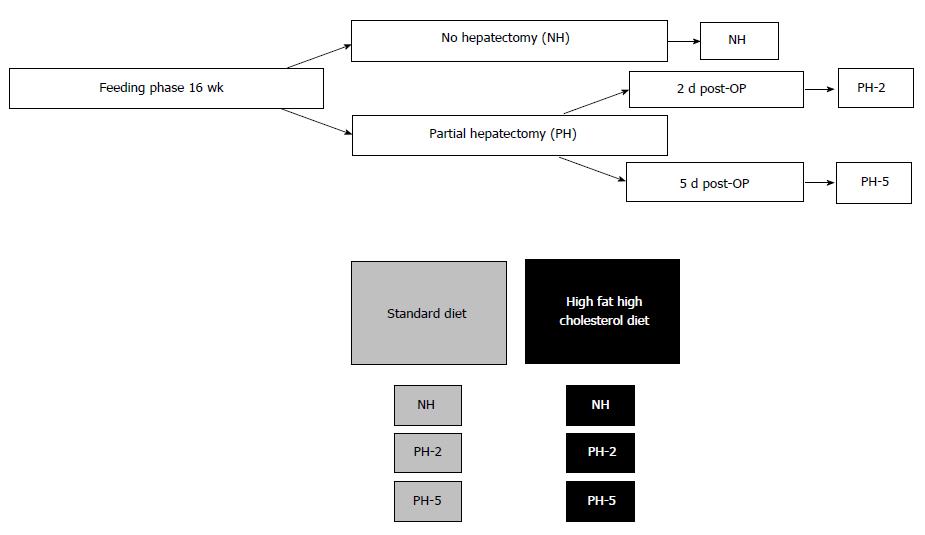
Figure 1 Overview of the groups, 9 animals in each group.
PH-2: Partial hepatectomy and 2 d; PH-5: Partial hepatectomy and 5 d.
Math 1 Math(A1).
Math 2 Math(A1).
Math 3 Math(A1).
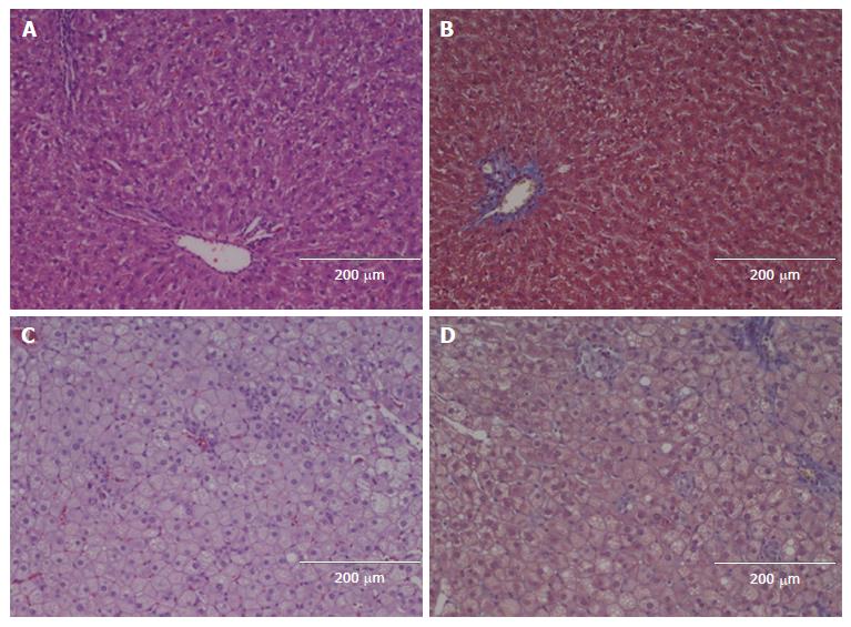
Figure 2 Examples of liver histology.
A and C shows Hematoxylin and Eosin; B and D shows Masson Trichome; A and B are from an animal fed the standard diet and euthanised without hepatectomy; C and D are from an animal fed the high fat high cholesterol diet and euthanised without hepatectomy.
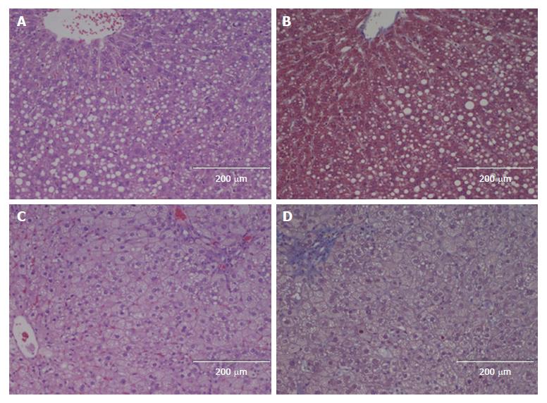
Figure 3 Examples of liver histology.
A and C shows Hematoxylin and Eosin; B and D shows Masson Trichome; A and B are from an animal fed the standard diet and euthanised two days after hepatectomy; C and D are from an animal fed the high fat high cholesterol diet and euthanised two days after hepatectomy.
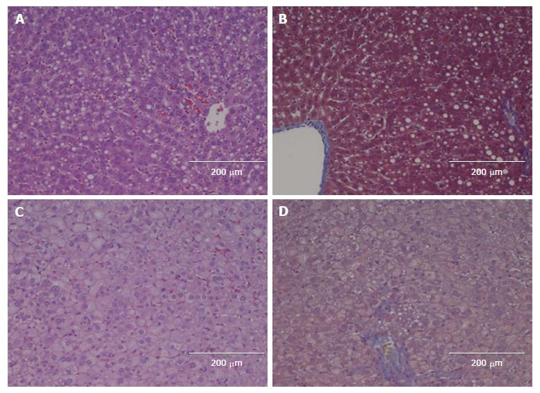
Figure 4 Examples of liver histology.
A and C shows Hematoxylin and Eosin; B and D shows Masson Trichome; A and B are from an animal fed the standard diet and euthanised five days after hepatectomy; C and D are from an animal fed the high fat high cholesterol diet and euthanised five days after hepatectomy.
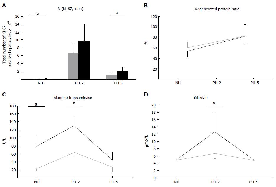
Figure 5 Total number of Ki-67 positive hepatocytes (A); regenerated protein ratio (B); Alanine transaminase (C) and Bilirubin (D).
Means and standard derivation displayed. aP < 0.05 compared to respective standard diet group. NH: No hepatectomy; PH-2: Partiel hepatectomy and 2 d; PH-5: Partiel hepatectomy and 5 d.
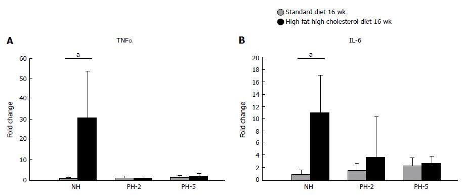
Figure 6 Relative mRNA expression.
A: Tumor necrosis factor α (TNFα); B: Interleukin-6 (IL-6) standardized to glyceraldehyde 3-phosphate dehydrogenase. Means and standard derivation displayed. Statistical analysis made using the ANOVA test, aP < 0.05 compared to respective standard diet group. NH: No hepatectomy; PH-2: Partiel hepatectomy and 2 d; PH-5: Partiel hepatectomy and 5 d.
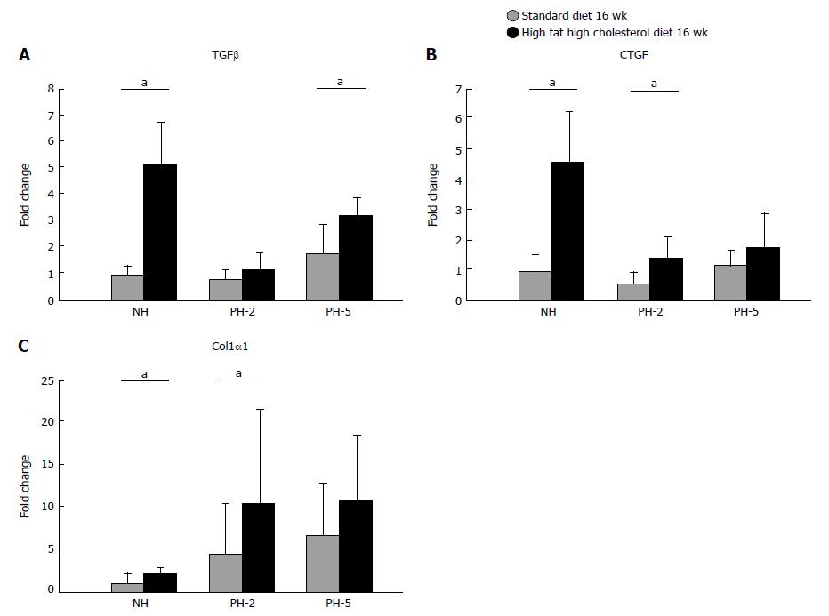
Figure 7 Relative mRNA expression.
A: Transforming Growth Factor β (TGFβ); B: Connective Tissue Growth Factor (CTGF); and C: Collagen 1α1 standardized (Col1α1) to glyceraldehyde 3-phosphate dehydrogenase (GAPDH). Means and standard derivation displayed. Statistical analysis made using the ANOVA test, aP < 0.05 compared to respective standard diet group. NH: No hepatectomy; PH-2: Partiel hepatectomy and 2 d; PH-5: Partiel hepatectomy and 5 d.
Figure 8 Relative mRNA expression.
A: Hepatocyte growth factor (HGF); B: Proto-oncogene, tyrosine kinase (MET); C: Epidermal growth factor (EGF); and D: Transforming growth factor α (TGFα), standardized to glyceraldehyde 3-phosphate dehydrogenase (GAPDH). Means and standard derivation displayed. Statistical analysis made using the ANOVA test, aP < 0.05 compared to respective standard diet group. NH: No hepatectomy; PH-2: Partiel hepatectomy and 2 d; PH-5: Partiel hepatectomy and 5 d.
- Citation: Haldrup D, Heebøll S, Thomsen KL, Andersen KJ, Meier M, Mortensen FV, Nyengaard JR, Hamilton-Dutoit S, Grønbæk H. Preserved liver regeneration capacity after partial hepatectomy in rats with non-alcoholic steatohepatitis. World J Hepatol 2018; 10(1): 8-21
- URL: https://www.wjgnet.com/1948-5182/full/v10/i1/8.htm
- DOI: https://dx.doi.org/10.4254/wjh.v10.i1.8


