
















修回日期: 2026-07-10
接受日期: 2026-07-27
在线出版日期: 2026-09-28
门静脉血栓(portal vein thrombosis, PVT)是肝硬化患者常见的并发症, 其发病率随着肝病进展及门静脉高压加重而增加. 门静脉血流动力学异常、凝血-纤溶系统失衡及肠道菌群紊乱等多种因素相互作用, 共同参与PVT形成. PVT形成可能加重腹水、增加食管胃底静脉曲张破裂出血风险, 并降低肝移植生存率. 抗凝治疗是症状性、完全闭塞性PVT、累及肠系膜上静脉者及肝移植候选者的核心干预手段, 而PVT形成可无明显临床表现, 且病程中存在自发再通或进展, 对抗凝治疗的时机带来挑战. 本文系统综述肝硬化PVT的临床特征、危险因素、抗凝药物及诊疗新靶点, 为临床规范化管理提供参考.
核心提要: 肝硬化门静脉血栓(portal vein thrombosis, PVT)形成是多因素介导的复杂病理生理过程, 需明确其自然病程与临床分型. 肝硬化患者抗凝治疗获益与出血风险增加之间权衡, 凸显了PVT诊疗决策的复杂与挑战. 抗凝治疗是特定患者的核心干预手段, 可改善临床结局, 新兴干预靶点的疗效与安全性仍待进一步评价.
引文著录: 靳珂珂, 丁惠国. 肝硬化门静脉血栓形成防治. 世界华人消化杂志 2026; 34(9): 675-684
Revised: July 10, 2026
Accepted: July 27, 2026
Published online: September 28, 2026
Portal vein thrombosis (PVT) is a common complication in patients with liver cirrhosis, and its incidence increases with the progression of liver disease and the aggravation of portal hypertension. The formation of PVT results from the interplay of multiple factors, including abnormal portal hemodynamics, coagulation-fibrinolysis imbalance, and intestinal flora dysbiosis. PVT may exacerbate ascites, increase the risk of esophagogastric variceal rupture and bleeding, and reduce the survival rate of liver transplantation recipients. Anticoagulant therapy is the core intervention for symptomatic, complete occlusive PVT, PVT extending to the superior mesenteric vein, and liver transplant candidates. However, PVT may be asymptomatic in its early stages, and its natural course is characterized by either spontaneous recanalization or progression, which poses challenges for the timing of anticoagulant therapy. This article systematically reviews the clinical characteristics, risk factors, anticoagulant agents, and novel diagnostic and therapeutic targets of PVT in cirrhotic patients, so as to provide a reference for the standardized clinical management of PVT.
- Citation: Jin KK, Ding HG. Prevention and treatment of portal vein thrombosis in cirrhosis. Shijie Huaren Xiaohua Zazhi 2026; 34(9): 675-684
- URL: https://www.wjgnet.com/1009-3079/full/v34/i9/675.htm
- DOI: https://dx.doi.org/10.11569/wcjd.v34.i9.675
核心提要: 肝硬化门静脉血栓(portal vein thrombosis, PVT)形成是多因素介导的复杂病理生理过程, 需明确其自然病程与临床分型. 肝硬化患者抗凝治疗获益与出血风险增加之间权衡, 凸显了PVT诊疗决策的复杂与挑战. 抗凝治疗是特定患者的核心干预手段, 可改善临床结局, 新兴干预靶点的疗效与安全性仍待进一步评价.
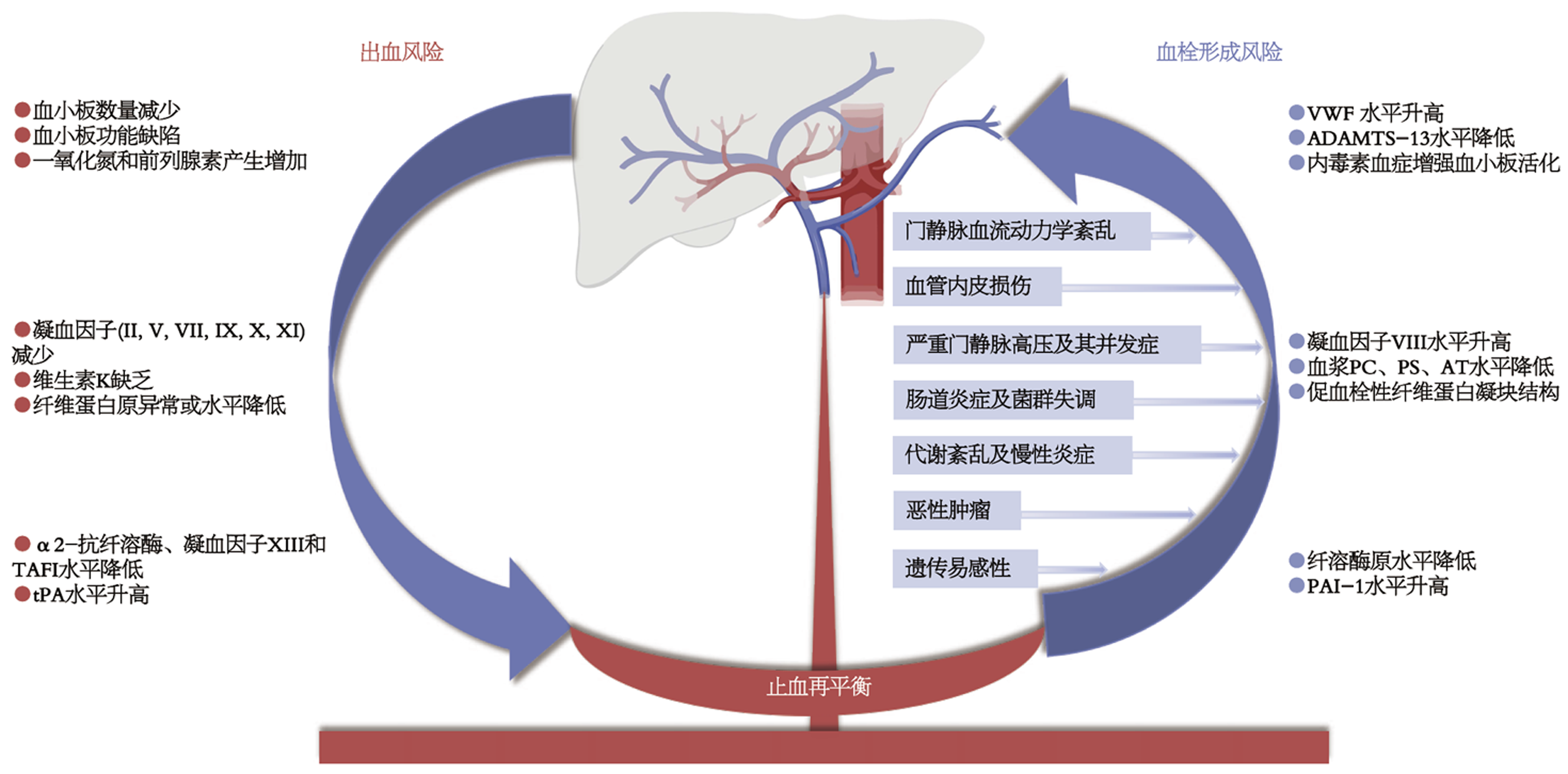
肝脏作为人体止血调控过程的核心场所, 参与多数凝血因子的合成与代谢. 肝硬化患者的止血系统由于促凝和抗凝同时改变而处于"止血再平衡状态"[1,2]. 在终末期肝病患者中, 这是一种脆弱的再平衡, 易受感染、肾功能损伤、酒精等因素影响而失衡, 进而导致出血或血栓形成[3-5]. 门静脉血栓(portal vein thrombosis, PVT)指门静脉系统内血栓形成, 包括门静脉主干及肝内门静脉分支, 可延伸至肠系膜上静脉或脾静脉[6]. PVT对肝硬化患者自然史的临床影响尚存在争议, 有研究表明[7-9], PVT形成可能加重门静脉高压, 增加食管胃底静脉曲张破裂出血风险, 降低肝癌患者生存率及肝移植成功率. PVT自然病程复杂, 存在自发再通、稳定及进展等不同转归[10], 其风险分层、治疗时机与方案选择仍面临困难与挑战.
PVT是肝硬化患者常见且严重的并发症, 年发病率为3%-17%[11]. PVT发病率与肝病严重程度相关, 在一项中位随访时间29(1-58) mo、Child A级肝硬化患者占76.3%的前瞻性队列[12], 1年、3年PVT累积发病率为3.7%、7.6%. 另一项荟萃分析发现[13], Child B级、C级肝硬化患者PVT发病率显著高于Child A级患者[18.34%(95%CI: 10.79%-27.35%) vs 9.89%(95%CI: 4.95%-16.30%), 亚组交互P<0.10]. 病毒性肝炎肝硬化患者1年、5年、8-10年PVT累积发病率为12.8%、20%、38.7%[14]; 非酒精性脂肪性肝炎患者PVT患病率高于其他病因肝硬化患者(10.1% vs 6.0%)[15].
肝硬化患者PVT可出现自发再通或进展, 临床结局事件与随访时间有关. 一项纳入26项研究[10](n = 1441)的荟萃分析发现, 约25%患者血栓出现进展, 45%保持稳定, 30%得到改善, 随访期间PVT复发率为24.0%(95%CI: 14.7-33.4). 研究发现[16], 直径<9 mm的自发性门体侧支分流(spontaneous portosystemic shunts, SPSS)可以独立预测PVT自发再通, 而粗大SPSS可产生血流窃血效应, 分流门静脉主干血流、加剧血流淤滞, 不利于血栓消退; 此外, 随访期间终末期肝病模型评分改善也与PVT再通有关[17]. 目前缺乏可靠模型预测PVT自发再通及对抗凝治疗应答人群. 因此, 对于PVT患者, 临床需标准化记录其初始部位、累及范围及闭塞程度, 以动态监测PVT转归及治疗应答情况.
肝硬化PVT的形成是多因素参与的复杂病理生理过程, 基于血栓形成的"Virchow三要素", 肝硬化背景下, PVT形成的机制可概述为门静脉血流速度减慢、血管内皮损伤和血液高凝状态等.
门静脉血流速度减慢、管腔扩张可造成血流瘀滞和涡流形成. 门静脉血流速度<15 cm/s是PVT发生的独立危险因素[危险比(hazard ratio, HR) = 2.66, 95%CI: 1.07-6.61, P = 0.035][18]. 长期应用非选择性β受体阻滞剂也可能是肝硬化患者PVT形成的危险因素[19]. 四维血流(4D Flow)磁共振成像(magnetic resonance imaging, MRI)检测发现, 门静脉相对滞留时间(relative residence time, RRT)延长亦可增加PVT发生风险[比值比(odds ratio, OR) = 11.4, 95%CI: 2.19-118, P = 0.001][20], 提示RRT延长可作为评估门静脉血流淤滞状态的新型预测指标. 门静脉增宽常见于肝硬化患者, 其直径≥14.5 mm可纳入PVT形成的危险因素预测模型[21].
脾切除/栓塞术、内镜下静脉曲张治疗可造成血管内皮机械性损伤, 激活凝血系统和血小板活化, 增加PVT形成风险[22,23]. Abdallah等[24]的研究发现, 肝硬化患者血小板数量减少, 但血小板聚集功能高于非PVT组, 提示血小板功能相对增强参与PVT形成. 此外, 体内血管性血友病因子裂解酶(A disintegrin and metalloproteinase with thrombospondin type 1 motifs, member 13, ADAMTS-13)活性降低, 血管性血友病因子(von Willebrand factor, vWF)蓄积, 导致血小板过度黏附聚集, 以18.8%为临界值, 低ADAMTS-13活性不仅是PVT形成独立危险因素, 还与患者不良预后密切相关; 凝血因子Ⅷ(factor Ⅷ, FⅧ)作为反映血管内皮损伤与高凝状态的指标, 在PVT患者中显著升高, 校正肝功能、脾切除等混杂因素后, FⅧ活性≥181.5%可独立预测基线无血栓肝硬化患者1年内新发PVT风险[25,26].
门静脉高压严重程度被认为是PVT形成的危险因素之一[27]. 研究发现[28], 肝静脉压力梯度(hepatic venous pressure gradient, HVPG)≥16 mmHg可作为PVT高危筛查阈值, 预测敏感性100.0%、特异性35.7%. 然而, 该阈值主要基于单中心小样本数据, 尚需前瞻性多中心大样本验证. 一项前瞻性研究(n = 369)表明[18], 所有新发PVT患者基线HVPG均>10 mmHg, 提示存在临床显著性门静脉高压; HVPG>20 mmHg患者PVT发生率显著升高(19% vs 2.4%, P = 0.015); 门静脉血流速度<15 cm/s、血小板减少、静脉曲张出血史均为PVT形成的独立危险因素. Driever等[29,30]发现, 肝硬化患者PVT以门静脉内膜纤维化增生为主, 仅约1/3的患者合并富纤维蛋白血栓, 这提示门静脉高压相关的血管重塑是PVT形成的重要机制之一.
肝硬化患者存在肠道菌群失调, 主要表现为有益菌丰度降低, 致病菌异常富集[31]. 研究发现[32,33], 肝硬化合并PVT患者肠道菌群多样性升高, 厌氧杆菌、假单胞菌等致病菌丰度明显增加, 这些致病菌引起的炎症状态和代谢产物失衡导致凝血级联激活, 可能介导PVT形成; 拟杆菌属丰度显著下降, 通过对四氯化碳诱导的肝硬化大鼠模型外源性补充拟杆菌, 可有效逆转PVT形成, 提示其具有潜在治疗价值. 此外, 肠道菌群失调可损伤肠道黏膜屏障、增加肠道通透性, 使革兰阴性菌裂解产生的脂多糖(lipopolysaccharide, LPS)大量易位至门静脉系统, 形成持续低水平内毒素血症, 而LPS通过激活Toll样受体4(toll-like receptor 4, TLR4)信号通路, 多途径促进PVT形成[34,35].
肝硬化合并PVT的荟萃分析发现[36], 糖尿病、NAFLD、高胆固醇血症可分别使肝硬化患者PVT风险增加1.80倍、1.61倍、3.59倍. 孟德尔随机化研究证实[37], 特定循环慢性炎症蛋白与PVT存在因果关联: 真核翻译起始因子4E结合蛋白1作为雷帕霉素靶蛋白信号通路下游的关键分子, 其水平升高可显著降低PVT风险(OR = 0.34, 95%CI: 0.15-0.79, P = 0.012), 而白细胞介素-6(lnterleukin-6, IL-6)等促炎因子则呈相反效应, 提示炎症信号网络的平衡状态影响PVT易感性.
肝细胞癌(hepatocellular carcinoma, HCC)可通过多种机制增加PVT形成风险[38]. 研究发现[39], HCC与肝硬化患者血小板聚集功能增强相关, 且该效应独立于血小板计数和肝硬化严重程度, HCC组vWF抗原水平显著高于非HCC组(348% vs 267%, P = 0.006). Zanetto等[40]的研究表明, HCC(HR = 10.34, 95%CI: 1.2-171.4, P = 0.03)及血栓弹力图参数-最大凝块硬度(maximum clot firmness, MCF)(HR = 6, 95%CI: 1-48, P = 0.001)均为PVT形成的独立危险因素, 其中MCF>25 mm可使PVT发生风险增加4.8倍.
遗传性易栓基因突变是肝硬化患者PVT形成的潜在危险因素. Amitrano等[41]研究发现, 凝血酶原基因G20210A突变(34.8% vs 2.5%)和MTHFR C677T纯合突变(43.5% vs 5.0%)在肝硬化合并PVT患者中显著升高, 69.5%的PVT患者可检出至少一种相关基因突变. Saugel等[42]研究指出, JAK2基因V617F突变在PVT患者中的检出率显著高于无PVT患者(10% vs 0%). 荟萃分析表明[43], 凝血因子V Leiden突变(OR = 1.98, 95%CI: 1.06-3.68, P = 0.03)与凝血酶原G20210A突变(OR = 2.43, 95%CI: 1.07-5.53, P = 0.03)均显著增加肝硬化患者PVT发生风险. 值得注意的是, PVT遗传易感因素存在种族差异, 《肝硬化门静脉血栓管理专家共识(2020年, 上海)》[6]指出, 遗传性易栓症并非中国肝硬化患者发生PVT的主要危险因素, 对脾大伴血小板正常或升高者, 建议筛查骨髓增殖性肿瘤相关基因以排除隐匿促栓因素. 肝硬化患者的止血再平衡及血栓形成风险见图1[2].
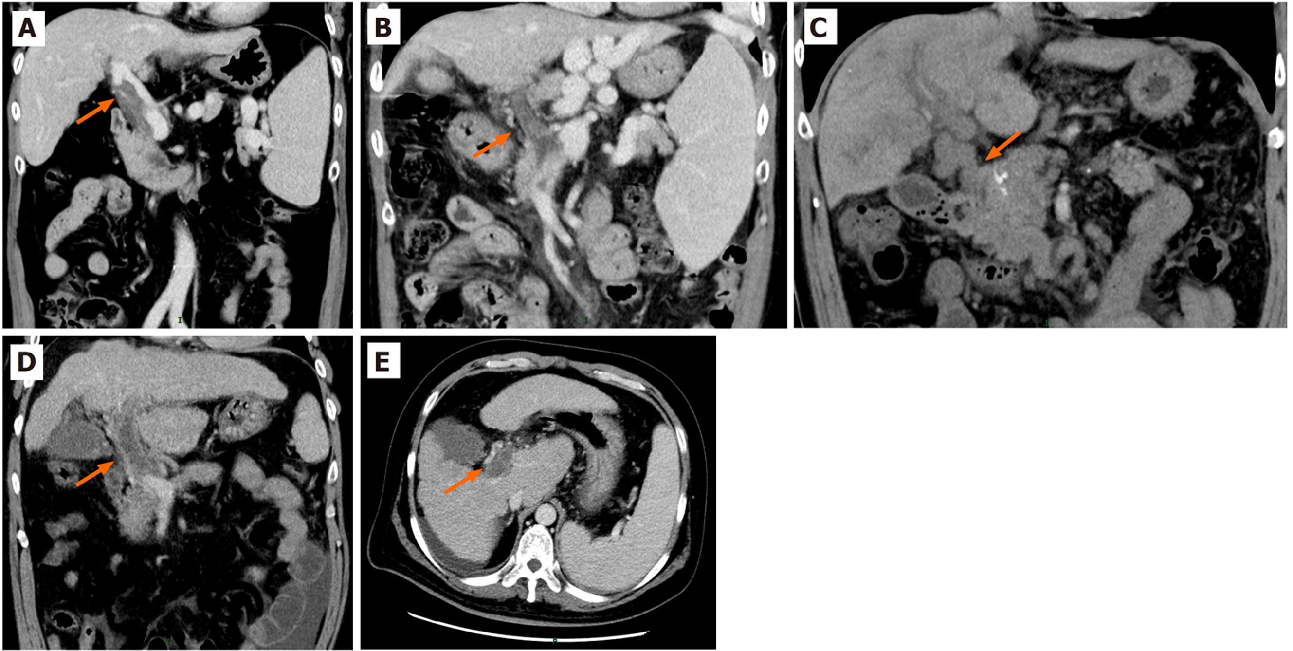
根据形成时间, PVT可分为急性(<6 mo)、慢性(>6 mo), 若慢性PVT范围扩大或累及肠系膜静脉、脾静脉, 出现腹痛、门静脉高压相关并发症加重, 甚至肠缺血坏死, 称为慢加急性PVT[44]. 基于影像学及病理特征的新分型, PVT可分为四类[45](见图2): 急性型表现为门静脉内极低密度均匀血栓, 无侧支循环及血管壁增厚, 病理以红细胞和血小板为主; 慢性型可见稍低密度均匀或不均匀血栓, 侧支循环增多伴血管壁增厚, 病理含纤维组织增生; 海绵样变型正常门静脉结构消失, 代以大量侧支血管, 病理以纤维组织和新生小血管增生为为主; 混合型则同时存在两种及以上分型表现, 病理呈层状混合血栓结构.
PVT可累及门静脉主干以及上游的肠系膜上静脉和脾静脉、下游的左右门静脉分支. 通过增强影像上垂直于门静脉主干平面计算门静脉主干残余管腔百分比(percentage of remnant lumen of the main portal vein, %RL), 可将PVT分为轻微阻塞(%RL≥50%)、部分阻塞(%RL<50%)和完全阻塞(%RL = 0%); 对于上、下游静脉, 可采用半定量量表进行视觉评估, 分为无血栓、不完全血栓(血栓可见残余管腔)和完全血栓(无残余管腔)[27]. 根据Yerdel分型[46], 将PVT分为4级: 1级: 门静脉管腔闭塞<50%, 伴或不伴肠系膜上静脉近端微小延伸; 2级: 门静脉管腔闭塞>50%, 伴或不伴肠系膜上静脉近端微小延伸; 3级: 门静脉及近端肠系膜上静脉完全闭塞, 远端肠系膜上静脉通畅; 4级: 门静脉、肠系膜上静脉近端及远端完全闭塞.
基于欧洲血管性肝病研究组(vascular liver disease interest group, VALDIG)标准[27], PVT演变可分为四类, (1)完全再通: 所有血栓均消失; (2)改善: 门静脉主干血栓完全消失, 且上、下游静脉血栓未完全消失, 无新发血栓, 或门静脉主干%RL增加超过25%, 且上、下游静脉血栓完全消失, 无新发血栓; (3)稳定: 门静脉主干%RL变化不超过25%, 且上、下游静脉血栓未完全消失或进展, 无新发血栓; 和(4)进展: 门静脉主干%RL减少超过25%, 或任意上、下游血管血栓进展, 或出现新发血栓.
肝硬化PVT治疗的目标: 尽早使血栓部分或完全再通, 且防止或减少血栓复发; 监测抗凝药物的安全性, 避免药物相关的严重不良事件.
肝硬化合并PVT的治疗包括抗凝、溶栓、血管介入治疗[47]. 抗凝治疗作为实现血管再通的一线核心措施, 不仅可以减少PVT形成, 还可在不显著增加出血风险的前提下, 降低腹水、肝性脑病的风险[48]. 鉴于肝硬化患者存在特殊的血栓与出血再平衡状态, 溶栓(全身或局部)在急性PVT(发病<14 d)中完全再通率较高, 但其出血风险很大, 除非有丰富临床经验的医生, 肝硬化PVT不建议常规溶栓治疗[49]. 血管介入治疗, 包括取血栓和经颈静脉肝内门体分流术, 仅适宜抗凝药物治疗无效的患者, 仍需要个体化综合评估.
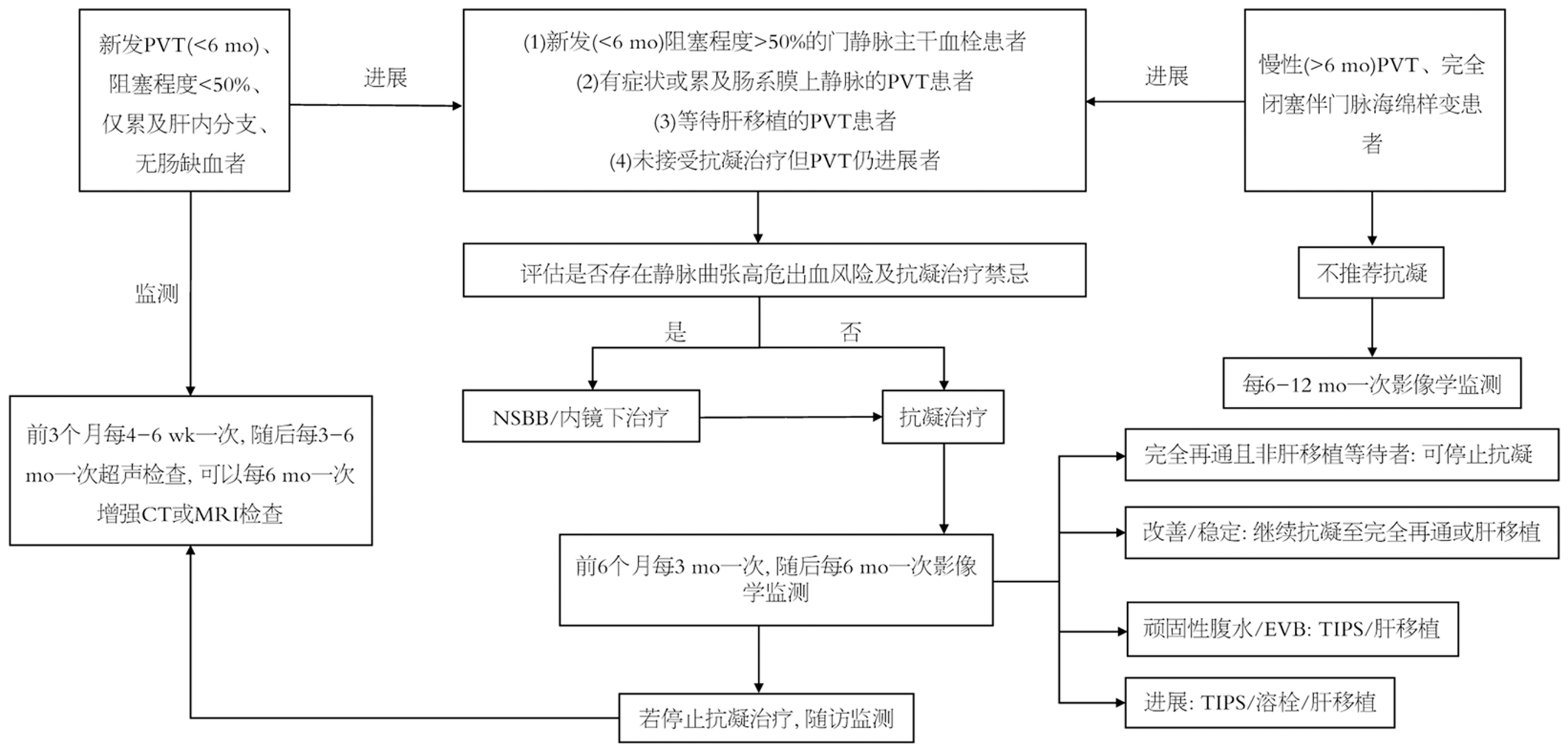
5.1.1 抗凝药物选择及疗程: 对于近期PVT形成, 尤其是合并肠系膜缺血, 以及潜在的肝移植候选者, 首选皮下注射低分子肝素, 后续可序贯维生素K拮抗剂或直接口服抗凝药(direct oral anticoagulants, DOACs)[50]. 尽管舒洛地特(口服类肝素制剂)在防治普通血栓性疾病中具有良好的安全性和有效性, 但尚缺乏治疗肝硬化PVT的高质量临床证据. 对于病程超过6 mo的慢性PVT、且血管完全闭塞并形成门静脉海绵样变性的患者, 血管再通概率较低, 不推荐常规抗凝治疗; 而慢性PVT仅部分闭塞、未形成海绵样变性的患者, 血管再通概率相对较高, 对于肝移植候选等潜在获益者, 仍可选择个体化抗凝治疗[51]. 肝硬化患者PVT的抗凝治疗药物详见表1[11,51-53].
| 药物 | 维生素K拮抗剂 | 低分子肝素 | 阿哌沙班 | 利伐沙班 | 艾多沙班 | 达比加群 |
| 作用机制 | 抑制维生素K依赖凝血因子(Ⅱ、Ⅶ、Ⅸ、Ⅹ) | 激活抗凝血酶 | Xa因子抑制剂 | Xa因子抑制剂 | Xa因子抑制剂 | IIa因子抑制剂 |
| 肝肾代谢 | 肝脏为主 | 肾脏: 40%; 肝脏: 60% | 肾脏: 27%; 肝脏: 73% | 肾脏: 66%; 肝脏: 34% | 肾脏: 50%; 肝脏: 50% | 肾脏: 80%; 肝脏: 20% |
| 标准剂量 | 基于INR值, 起始剂量2.5-5 mg/日, 目标INR: 2.0-3.0 | 按体重给药: 依诺肝素100 IU/kg/次/12 h或150 IU/kg/次/日; 那曲肝素钙100 IU/kg/次/12 h; 达肝素钠100 IU/kg/次/12 h或200 IU/kg/日 | 2.5 mg, 每日2次(数据有限) | 临床证据有限, 一般个体化使用剂量10-20 mg/日 | 60 mg, 每日1次; <60 kg或CrCl 15-50 mL/min, 30 mg, 每日1次 | 150 mg, 每日2次; 年龄≥80岁、合用钙通道阻滞剂或其他高出血风险人群, 调整为110 mg, 每日2次 |
| 按Child-Pugh分级调整用药 | ||||||
| Child A | 标准剂量 | 标准剂量 | 标准剂量 | 无推荐剂量 | 标准剂量 | 标准剂量 |
| Child B | 标准剂量 | 标准剂量 | 慎用 | 不推荐使用 | 不推荐使用 | 慎用 |
| Child C | 标准剂量 | 标准剂量 | 不推荐使用 | 不推荐使用 | 不推荐使用 | 不推荐使用 |
| 按肾功能不全调整用药 | ||||||
| CrCl 30-50 mL/min | 无需调整剂量 | 无需调整剂量 | 无需调整剂量 | 减量 | 减量 | 无需调整剂量 |
| CrCl 15-30 mL/min | 无需调整剂量 | 减量 | 慎用 | 不推荐使用 | 减量 | 不推荐使用 |
| 拮抗剂 | 维生素K、4F-PCC | 硫酸鱼精蛋白 | Andexanet alfa、4F-PCC | Andexanet alfa、4F-PCC | 4F-PCC、Andexanet alfa(超说明书) | Idarucizumab |
| 优点 | 价格低廉, 口服给药; CrCl<15 mL/min可用 | 循证证据充分; 半衰期短. 可固定剂量. 舒洛地特(口服类肝素制剂)尚无证据 | 口服给药 | 口服给药 | 口服给药 | 口服给药 |
| 缺点 | 常规监测INR; 严重肝病出血风险高 | 皮下注射; 需按体重给药. CrCl<15 mL/min禁用 | CrCl<15 mL/min禁用1 | CrCl<30 mL/min不推荐使用 | CrCl<15 mL/min禁用 | ALT>2×ULN时不推荐使用1 |
抗凝治疗启动后3 mo行增强计算机断层扫描(computed tomography, CT)评估疗效, 若门静脉再通, 疗程至少维持6 mo; 肝移植候选者需持续抗凝直至移植手术前; 其他患者在再通后应权衡预防血栓复发、生存获益与出血风险, 个体化评估是否继续抗凝, 并建议每3 mo复查影像学; 依从性良好但抗凝治疗无应答、血栓仍进展的患者, 可考虑行补救性血管内介入治疗或停用抗凝药物[51]. 值得注意的是, 门静脉再通及抗凝治疗终止后, 仍有高达38%的患者可在2-5 mo内出现PVT复发[54].
5.1.2 抗凝治疗监测: 在PVT的全程管理中, 随访监测与出血事件处理是改善预后、降低并发症的关键环节: (1)凝血指标: 凝血时间、凝血酶原时间等常规凝血指标, 不能用于评估抗凝疗效或预测出血风险[55]. 抗凝治疗1 wk, 血浆纤维蛋白降解产物(fibrin degradation product, FDP)和D-二聚体显著下降, 认为抗凝药物治疗有效. 但是, FDP和D-二聚体作为判断评价疗效的指标需要进一步研究. 以血栓弹力图为代表的全程凝血功能检测, 具有良好应用潜力, 但仍需更多研究验证[56]; 和(2)影像学检查与频率: 根据VALDIG建议, 多普勒超声可作为一线筛查手段, 增强CT/MRI则用于明确诊断及疗效评价的金标准, 测量%RL以量化血栓负荷及动态演变[27]. 肝硬化PVT患者抗凝治疗的管理及监测详见图3[6,27].
5.1.3 出血风险管理: 血小板计数<50×109/L对肝硬化患者抗凝治疗出血风险的预测价值有限[54]. Zanetto等[57]发现血栓弹力图最大振幅(maximum amplitude, MA)在预测操作相关出血方面优于血小板计数, MA<30 mm可识别高危出血患者. 作为综合评估血小板功能与血凝块强度的指标, MA指导个体化抗凝治疗仍需要进一步研究. 现有证据表明[58], 内镜下静脉曲张套扎术可以在不停用抗凝药物的情况下进行, 且不增加静脉曲张出血风险. 一项荟萃分析发现[59], 抗凝治疗可显著降低患者全因死亡率(调整后的亚分布危险比 = 0.59, 95%CI: 0.49-0.70, P<0.001), 该获益独立于PVT的再通和严重程度, 但增加非门脉高压性出血风险(9.7% vs 1.7%, P<0.001).
欧洲肝脏研究学会指南[60]建议, 行侵入性操作前, 抗凝药物应遵循与无肝硬化患者相同的指南管理. 对于抗凝期间发生的出血事件, 首先根据出血严重程度分层处理, 轻度出血以局部措施为主, 可暂缓单次用药, 无需长期停用抗凝, 不推荐常规输注血液制品; 中-重度出血需暂停抗凝治疗, 并积极处理感染、肾功能不全等加重出血的因素, 对服用DOACs的患者可监测其血药浓度, 若浓度≥50 ng/mL, 提示其抗凝活性显著并增加出血风险, 可个体化选择相应拮抗剂; 输注凝血酶原复合物浓缩物需评估血栓形成风险[61,62]; 对于门脉高压相关出血, 应优先选用降低门脉压力药物或内镜治疗控制出血, 仅在一线治疗无效时考虑纠正凝血异常[60].
出血控制后建议个体化评估重启抗凝, 但重启抗凝时机尚未形成统一标准. 一项多中心随机对照研究证实[63], 肝硬化合并急性静脉曲张破裂出血且伴PVT的患者, 在食管静脉曲张套扎术后48 h内启动那屈肝素-华法林序贯抗凝可提高PVT再通率(67.4% vs 39.5%, P = 0.009), 且不增加术后再出血风险. 反复静脉曲张出血者可考虑门静脉再通术-经颈静脉肝内门体分流术(portal vein revascularizationtransjugular intrahepatic portosystemic shunt, PVR-TIPS)[51].
肠道菌群失调、肠道屏障损伤、慢性炎症及代谢紊乱可共同驱动肝硬化PVT发生, 成为传统抗凝治疗之外的重要干预方向.
5.2.1 肠道菌群与黏膜屏障: 肠肝循环受损是肠道菌群紊乱和黏膜屏障损伤的重要原因. 一项对比健康人群和肝病患者肠道菌群的研究发现[64], 肝病患者肠道双歧杆菌属及拟杆菌属丰度降低, 而肠杆菌属与变形杆菌门则显著增加; 小肠细菌过度生长主要由胆汁分泌不足与胃肠蠕动延迟共同驱动, 可进一步导致肠道屏障通透性受损. 因此, 靶向调节肠道微生物群(如粪菌移植或靶向抗生素)可作为潜在治疗方式[65]. 法尼醇X受体(farnesoid X receptor, FXR)作为胆汁酸代谢关键核受体, 参与调控肠道屏障结构与功能、抑制细菌易位. 肝硬化患者肠道胆汁酸缺乏可导致FXR通路下调, 加剧屏障破坏, 而FXR激动剂可修复肠道多重屏障、减少细菌易位, 成为调控肝硬化患者肠-肝轴紊乱、防治PVT的潜在靶向药物[66].
5.2.2 慢性炎症与白蛋白: 短链脂肪酸作为肠道菌群发酵膳食纤维产生的关键代谢产物, 可通过上调紧密连接蛋白表达强化肠道屏障功能, 抑制菌群易位与炎症反应, 对肠道屏障修复具有作用[67,68]. 针对LPS介导门静脉系统的促炎及促血栓机制, 降低循环LPS水平或拮抗其毒性是降低PVT风险的重要方式. 肠道碱性磷酸酶可修饰LPS脂质A结构, 使其去磷酸化转化为拮抗型单磷酸化LPS, 竞争性结合TLR4, 从源头上降低LPS负荷及促栓活性, 为PVT防治提供新靶点[69]. 此外, 机体可通过白蛋白中和LPS发挥天然防御作用, 而肝硬化患者低白蛋白血症, 这不仅会削弱机体对LPS的解毒能力, 还会因白蛋白本身具备的抗血小板作用受损而增加血栓形成风险[70]. 因此, 靶向降低内毒素血症及补充白蛋白, 可以从控制炎症、减轻凝血激活两方面协同降低PVT发生风险.
PVT是肝硬化患者常见的并发症, 可能加重腹水、食管胃底静脉曲张破裂出血等门脉高压并发症, 并降低肝移植生存率. 抗凝治疗是症状性、完全闭塞性PVT、累及肠系膜上静脉者及肝移植候选者的核心干预手段, 可改善患者的临床结局. 但是, 失代偿期肝硬化合并PVT的临床诊疗仍存在不足, 如何在抗凝治疗获益与终末期肝硬化患者出血风险增加之间权衡, 凸显了诊疗决策的复杂与挑战. 目前, 肝硬化PVT抗凝治疗的最佳启动时机、疗程尚无统一共识. 新型口服抗凝药物与各类新干预靶点的疗效和安全性及用于PVT风险分层、疗效监测及预后评价的生物标志物仍需要进一步研究.
| 1. | Bozic D, Babic A, Olic I, Lalovac M, Mijic M, Madir A, Podrug K, Mestrovic A. Coagulation Abnormalities in Liver Cirrhosis: Diagnostic and Therapeutic Approaches. Medicina (Kaunas). 2026;62:104. [PubMed] [DOI] |
| 2. | Lisman T, Caldwell SH, Intagliata NM. Haemostatic alterations and management of haemostasis in patients with cirrhosis. J Hepatol. 2022;76:1291-1305. [PubMed] [DOI] |
| 3. | Singh AD, Mucha SR, Lindenmeyer CC. Cirrhotic coagulopathy: A rebalanced hemostasis. Cleve Clin J Med. 2022;89:523-533. [PubMed] [DOI] |
| 4. | O'Leary JG, Greenberg CS, Patton HM, Caldwell SH. AGA Clinical Practice Update: Coagulation in Cirrhosis. Gastroenterology. 2019;157:34-43.e1. [PubMed] [DOI] |
| 5. | Howard BM, Kornblith LZ, Redick BJ, Conroy AS, Nelson MF, Calfee CS, Callcut RA, Cohen MJ. Exposing the bidirectional effects of alcohol on coagulation in trauma: Impaired clot formation and decreased fibrinolysis in rotational thromboelastometry. J Trauma Acute Care Surg. 2018;84:97-103. [PubMed] [DOI] |
| 7. | Zhang Y, Xu BY, Wang XB, Zheng X, Huang Y, Chen J, Meng ZJ, Gao YH, Qian ZP, Liu F, Lu XB, Shi Y, Shang J, Li H, Wang SY, Yin S, Sun SN, Hou YX, Xiong Y, Chen J, Li BL, Lei Q, Gao N, Ji LJ, Li J, Jie FR, Zhao RH, Liu JP, Lin TF, Chen LY, Tan WT, Zhang Q, Zou CC, Huang ZB, Jiang XH, Luo S, Liu CY, Zhang YY, Li T, Ren HT, Wang SJ, Deng GH, Xiong SE, Liu XX, Wang C, Yuan W, Gu WY, Qiao L, Wang TY, Wu DD, Dong FC, Li H, Hua J. Prevalence and Clinical Significance of Portal Vein Thrombosis in Patients With Cirrhosis and Acute Decompensation. Clin Gastroenterol Hepatol. 2020;18:2564-2572.e1. [PubMed] [DOI] |
| 8. | Senzolo M, Shalaby S, Grasso M, Vitale A, Pizzirani E, Barbiero G, Zanetto A, Feltracco P, Simioni P, Burra P, Cillo U. Role of nonneoplastic PVT in the natural history of patients with cirrhosis and first diagnosis of HCC. Hepatology. 2024;79:355-367. [PubMed] [DOI] |
| 9. | Rizzari MD, Safwan M, Sobolic M, Kitajima T, Collins K, Yoshida A, Abouljoud M, Nagai S. The Impact of Portal Vein Thrombosis on Liver Transplant Outcomes: Does Grade or Flow Rate Matter? Transplantation. 2021;105:363-371. [PubMed] [DOI] |
| 10. | Giri S, Singh A, Kolhe K, Kale A, Shukla A. Natural history of portal vein thrombosis in cirrhosis: A systematic review with meta-analysis. J Gastroenterol Hepatol. 2023;38:1710-1717. [PubMed] [DOI] |
| 11. | Meena BL, Sarin SK. Management of Portal vein Thrombosis in Cirrhosis. Semin Liver Dis. 2024;44:416-429. [PubMed] [DOI] |
| 12. | Noronha Ferreira C, Marinho RT, Cortez-Pinto H, Ferreira P, Dias MS, Vasconcelos M, Alexandrino P, Serejo F, Pedro AJ, Gonçalves A, Palma S, Leite I, Reis D, Damião F, Valente A, Xavier Brito L, Baldaia C, Fatela N, Ramalho F, Velosa J. Incidence, predictive factors and clinical significance of development of portal vein thrombosis in cirrhosis: A prospective study. Liver Int. 2019;39:1459-1467. [PubMed] [DOI] |
| 13. | Pan J, Wang L, Gao F, An Y, Yin Y, Guo X, Nery FG, Yoshida EM, Qi X. Epidemiology of portal vein thrombosis in liver cirrhosis: A systematic review and meta-analysis. Eur J Intern Med. 2022;104:21-32. [PubMed] [DOI] |
| 14. | Maruyama H, Okugawa H, Takahashi M, Yokosuka O. De novo portal vein thrombosis in virus-related cirrhosis: predictive factors and long-term outcomes. Am J Gastroenterol. 2013;108:568-574. [PubMed] [DOI] |
| 15. | Stine JG, Shah NL, Argo CK, Pelletier SJ, Caldwell SH, Northup PG. Increased risk of portal vein thrombosis in patients with cirrhosis due to nonalcoholic steatohepatitis. Liver Transpl. 2015;21:1016-1021. [PubMed] [DOI] |
| 16. | Yokoyama S, Ishizu Y, Honda T, Imai N, Ito T, Yamamoto K, Tsuzuki T, Ishigami M. Absence of large portal collateral vessels is associated with spontaneous improvement of cirrhotic portal vein thrombosis. Hepatol Res. 2022;52:199-209. [PubMed] [DOI] |
| 17. | Xu S, Guo X, Xu X, Wang L, Tacke F, Primignani M, He Y, Yin Y, Yi F, Qi X. Natural history and predictors associated with the evolution of portal venous system thrombosis in liver cirrhosis. Eur J Gastroenterol Hepatol. 2021;33:e423-e430. [PubMed] [DOI] |
| 18. | Turon F, Driever EG, Baiges A, Cerda E, García-Criado Á, Gilabert R, Bru C, Berzigotti A, Nuñez I, Orts L, Reverter JC, Magaz M, Camprecios G, Olivas P, Betancourt-Sanchez F, Perez-Campuzano V, Blasi A, Seijo S, Reverter E, Bosch J, Borràs R, Hernandez-Gea V, Lisman T, Garcia-Pagan JC. Predicting portal thrombosis in cirrhosis: A prospective study of clinical, ultrasonographic and hemostatic factors. J Hepatol. 2021;75:1367-1376. [PubMed] [DOI] |
| 19. | Nery F, Correia S, Macedo C, Gandara J, Lopes V, Valadares D, Ferreira S, Oliveira J, Gomes MT, Lucas R, Rautou PE, Miranda HP, Valla D. Nonselective beta-blockers and the risk of portal vein thrombosis in patients with cirrhosis: results of a prospective longitudinal study. Aliment Pharmacol Ther. 2019;49:582-588. [PubMed] [DOI] |
| 20. | Hyodo R, Takehara Y, Ishizu Y, Nishida K, Mizuno T, Ichikawa K, Horiguchi R, Kurata N, Ogura Y, Yokoyama S, Naganawa S, Jin N, Ichiba Y. Evaluation of 4D Flow MRI-Derived Relative Residence Time as a Marker for Cirrhosis Associated Portal Vein Thrombosis. J Magn Reson Imaging. 2024;60:2592-2601. [PubMed] [DOI] |
| 21. | Nie GL, Yan J, Li Y, Zhang HL, Xie DN, Zhu XW, Li X. Predictive model for non-malignant portal vein thrombosis associated with cirrhosis based on inflammatory biomarkers. World J Gastrointest Oncol. 2024;16:1213-1226. [PubMed] [DOI] |
| 22. | Xu X, Jin J, Liu Y, Li H. Analysis of related factors of portal vein thrombosis in liver cirrhosis. BMC Gastroenterol. 2023;23:26. [PubMed] [DOI] |
| 23. | Wang L, Guo X, Xu X, Philips CA, Primignani M, Mendez-Sanchez N, Li Q, Zheng K, Qi X. Association of portal venous system thrombosis with endoscopic variceal treatment: a systematic review and meta-analysis. Eur J Gastroenterol Hepatol. 2021;32:125-131. [PubMed] [DOI] |
| 24. | Abdallah HM, Elbassal F, Saber EM, Sabry A, Hendy OM, Nassar MR, Al-Morshedy SM. Platelet aggregation as a thrombotic marker in cirrhotic patients with portal vein thrombosis. Clin Exp Hepatol. 2025;11:179-189. [PubMed] [DOI] |
| 25. | Suzuki J, Namisaki T, Takya H, Kaji K, Nishimura N, Shibamoto A, Asada S, Kubo T, Iwai S, Tomooka F, Takeda S, Koizumi A, Tanaka M, Matsuda T, Inoue T, Fujimoto Y, Tsuji Y, Fujinaga Y, Sato S, Kitagawa K, Kawaratani H, Akahane T, Mitoro A, Matsumoto M, Asada K, Yoshiji H. ADAMTS-13: A Prognostic Biomarker for Portal Vein Thrombosis in Japanese Patients with Liver Cirrhosis. Int J Mol Sci. 2024;25:2678. [PubMed] [DOI] |
| 26. | Jiang S, Ai Y, Fan X, Huang X, Wu L, Ni L, Li F, Chen S. Increased Factor VIII Activity Is Predictive of the Occurrence of Portal Vein Thrombosis in Cirrhosis. Thromb Haemost. 2023;123:714-722. [PubMed] [DOI] |
| 27. | Elkrief L, Hernandez-Gea V, Senzolo M, Albillos A, Baiges A, Berzigotti A, Bureau C, Murad SD, De Gottardi A, Durand F, Garcia-Pagan JC, Lisman T, Mandorfer M, McLin V, Moga L, Nery F, Northup P, Nuzzo A, Paradis V, Patch D, Payancé A, Plaforet V, Plessier A, Poisson J, Roberts L, Salem R, Sarin S, Shukla A, Toso C, Tripathi D, Valla D, Ronot M, Rautou PE; ERN RARE-LIVER and VALDIG, an EASL consortium. Portal vein thrombosis: diagnosis, management, and endpoints for future clinical studies. Lancet Gastroenterol Hepatol. 2024;9:859-883. [PubMed] [DOI] |
| 28. | Zhong H, Lu S, Xu M, Liu N, Ye W, Yang Y. Predictive value of hepatic venous pressure gradient in cirrhotic portal vein thrombosis development. Clin Res Hepatol Gastroenterol. 2024;48:102484. [PubMed] [DOI] |
| 29. | Driever EG, von Meijenfeldt FA, Adelmeijer J, de Haas RJ, van den Heuvel MC, Nagasami C, Weisel JW, Fondevila C, Porte RJ, Blasi A, Heaton N, Gregory S, Kane P, Bernal W, Zen Y, Lisman T. Nonmalignant portal vein thrombi in patients with cirrhosis consist of intimal fibrosis with or without a fibrin-rich thrombus. Hepatology. 2022;75:898-911. [PubMed] [DOI] |
| 30. | Driever EG, Cannegieter SC, Gregory S, Kane P, Zen Y, Lisman T, Bernal W. Portal vein intimal thickening in patients with cirrhosis is associated with indicators of portal hypertension. J Thromb Haemost. 2025;23:1551-1561. [PubMed] [DOI] |
| 31. | Trebicka J, Bork P, Krag A, Arumugam M. Utilizing the gut microbiome in decompensated cirrhosis and acute-on-chronic liver failure. Nat Rev Gastroenterol Hepatol. 2021;18:167-180. [PubMed] [DOI] |
| 32. | Huang XY, Zhang YH, Yi SY, Lei L, Ma T, Huang R, Yang L, Li ZM, Zhang D. Potential contribution of the gut microbiota to the development of portal vein thrombosis in liver cirrhosis. Front Microbiol. 2023;14:1217338. [PubMed] [DOI] |
| 33. | Hasan RA, Koh AY, Zia A. The gut microbiome and thromboembolism. Thromb Res. 2020;189:77-87. [PubMed] [DOI] |
| 34. | Violi F, Pignatelli P, Castellani V, Carnevale R, Cammisotto V. Gut dysbiosis, endotoxemia and clotting activation: A dangerous trio for portal vein thrombosis in cirrhosis. Blood Rev. 2023;57:100998. [PubMed] [DOI] |
| 35. | Li W, Deng M, Loughran PA, Yang M, Lin M, Yang C, Gao W, Jin S, Li S, Cai J, Lu B, Billiar TR, Scott MJ. LPS Induces Active HMGB1 Release From Hepatocytes Into Exosomes Through the Coordinated Activities of TLR4 and Caspase-11/GSDMD Signaling. Front Immunol. 2020;11:229. [PubMed] [DOI] |
| 36. | Li J, Wang Q, Yang M, Sun X. Metabolic Disorders and Risk of Portal Vein Thrombosis in Liver Cirrhosis: A Systematic Review and Meta-Analysis. Turk J Gastroenterol. 2022;33:541-553. [PubMed] [DOI] |
| 37. | Zhang B, Yao Z, Li P, Niu G, Yan Z, She K, Cheng G, Yang M. Causal Relationships of Circulating Inflammatory Proteins and Portal Vein Thrombosis: A Mendelian Randomization Study. Semin Thromb Hemost. 2025;51:272-278. [PubMed] [DOI] |
| 38. | Stella L, De Siati M, Talerico R, Pallozzi M, Cerrito L, Sorrentino S, Gasbarrini A, De Candia E, Pola R, Ponziani FR. Thrombotic Risk and Coagulation Imbalance in Cirrhosis and Hepatocellular Carcinoma: Clinical Implications and Management. Cancers (Basel). 2025;17:3413. [PubMed] [DOI] |
| 39. | Zanetto A, Senzolo M, Campello E, Bulato C, Gavasso S, Shalaby S, Gambato M, Vitale A, Cillo U, Farinati F, Russo FP, Simioni P, Burra P. Influence of Hepatocellular Carcinoma on Platelet Aggregation in Cirrhosis. Cancers (Basel). 2021;13:1150. [PubMed] [DOI] |
| 40. | Zanetto A, Senzolo M, Vitale A, Cillo U, Radu C, Sartorello F, Spiezia L, Campello E, Rodriguez-Castro K, Ferrarese A, Farinati F, Burra P, Simioni P. Thromboelastometry hypercoagulable profiles and portal vein thrombosis in cirrhotic patients with hepatocellular carcinoma. Dig Liver Dis. 2017;49:440-445. [PubMed] [DOI] |
| 41. | Amitrano L, Brancaccio V, Guardascione MA, Margaglione M, Iannaccone L, D'Andrea G, Marmo R, Ames PR, Balzano A. Inherited coagulation disorders in cirrhotic patients with portal vein thrombosis. Hepatology. 2000;31:345-348. [PubMed] [DOI] |
| 42. | Saugel B, Lee M, Feichtinger S, Hapfelmeier A, Schmid RM, Siveke JT. Thrombophilic factor analysis in cirrhotic patients with portal vein thrombosis. J Thromb Thrombolysis. 2015;40:54-60. [PubMed] [DOI] |
| 43. | Ma SD, Wang J, Bezinover D, Kadry Z, Northup PG, Stine JG. Inherited thrombophilia and portal vein thrombosis in cirrhosis: A systematic review and meta-analysis. Res Pract Thromb Haemost. 2019;3:658-667. [PubMed] [DOI] |
| 45. | Lv YF, Liu BW, Han Y, Meng MM, Li DZ, Tian H, Wang FC, Dong JH, Yang YP, Zhou GD, Ding HG, Zhang YN, Liu FQ, Zhu B. Proposal of a new classification scheme for complete portal vein thrombosis and its clinical significance: A retrospective study. World J Gastroenterol. 2026;32:117479. [DOI] |
| 46. | Yerdel MA, Gunson B, Mirza D, Karayalçin K, Olliff S, Buckels J, Mayer D, McMaster P, Pirenne J. Portal vein thrombosis in adults undergoing liver transplantation: risk factors, screening, management, and outcome. Transplantation. 2000;69:1873-1881. [PubMed] [DOI] |
| 47. | Wu J, Deng X, Luo J, Jiang Z, Xie F, Chen B, Leung HW, Zhang G, To KF, Kang W. Update in the treatment of cirrhotic patients with portal vein thrombosis. Clin Mol Hepatol. 2025;31:1139-1166. [PubMed] [DOI] |
| 48. | Sato A, Watanabe S, Iseki M, Yamada Y, Kobayashi R, Furuya M, Arai K, Ohshina E, Tashiro Y, Nozaka T, Matsuoka M, Yauchi M, Kobayashi K, Matsumoto T, Furumoto Y, Asano T, Azuma S. Anticoagulation against portal vein thrombosis reduces mortality and liver cirrhosis-related complications: A propensity score-matched study. Hepatol Res. 2023;53:1096-1104. [PubMed] [DOI] |
| 49. | Primignani M, Tosetti G, Ierardi AM. Approach to different thrombolysis techniques and timing of thrombolysis in the management of portal vein thrombosis in cirrhotic patients. J Transl Int Med. 2023;11:198-202. [PubMed] [DOI] |
| 50. | de Franchis R, Bosch J, Garcia-Tsao G, Reiberger T, Ripoll C; Baveno VII Faculty. Baveno VII - Renewing consensus in portal hypertension. J Hepatol. 2022;76:959-974. [PubMed] [DOI] |
| 51. | Davis JPE, Lim JK, Francis FF, Ahn J. AGA Clinical Practice Update on Management of Portal Vein Thrombosis in Patients With Cirrhosis: Expert Review. Gastroenterology. 2025;168:396-404.e1. [PubMed] [DOI] |
| 52. | Al Naamani KM, Rizwan S, Al Sinani S, Al Rawahi B, AlMa'mari S, El-Kassas M. Direct Oral Anticoagulants for Portal Vein Thrombosis in Cirrhosis: From Pharmacology to Clinical Practice. Clin Appl Thromb Hemost. 2026;32:10760296261420229. [PubMed] [DOI] |
| 53. | Pereira Portela C, Gautier LA, Zermatten MG, Fraga M, Moradpour D, Bertaggia Calderara D, Aliotta A, Veuthey L, De Gottardi A, Stirnimann G, Alberio L. Direct oral anticoagulants in cirrhosis: Rationale and current evidence. JHEP Rep. 2024;6:101116. [PubMed] [DOI] |
| 54. | Delgado MG, Seijo S, Yepes I, Achécar L, Catalina MV, García-Criado A, Abraldes JG, de la Peña J, Bañares R, Albillos A, Bosch J, García-Pagán JC. Efficacy and safety of anticoagulation on patients with cirrhosis and portal vein thrombosis. Clin Gastroenterol Hepatol. 2012;10:776-783. [PubMed] [DOI] |
| 55. | Zermatten MG, Fraga M, Calderara DB, Aliotta A, Moradpour D, Alberio L. Biomarkers of liver dysfunction correlate with a prothrombotic and not with a prohaemorrhagic profile in patients with cirrhosis. JHEP Rep. 2020;2:100120. [PubMed] [DOI] |
| 56. | Tripodi A, Primignani M, Mannucci PM, Caldwell SH. Changing Concepts of Cirrhotic Coagulopathy. Am J Gastroenterol. 2017;112:274-281. [PubMed] [DOI] |
| 57. | Zanetto A, Rinder HM, Senzolo M, Simioni P, Garcia-Tsao G. Reduced Clot Stability by Thromboelastography as a Potential Indicator of Procedure-Related Bleeding in Decompensated Cirrhosis. Hepatol Commun. 2021;5:272-282. [PubMed] [DOI] |
| 58. | Bianchini M, Cavani G, Bonaccorso A, Turco L, Vizzutti F, Sartini A, Gitto S, Merighi A, Banchelli F, Villa E, Schepis F. Low molecular weight heparin does not increase bleeding and mortality post-endoscopic variceal band ligation in cirrhotic patients. Liver Int. 2018;38:1253-1262. [PubMed] [DOI] |
| 59. | Guerrero A, Campo LD, Piscaglia F, Scheiner B, Han G, Violi F, Ferreira CN, Téllez L, Reiberger T, Basili S, Zamora J, Albillos A; Baveno Cooperation: an EASL consortium. Anticoagulation improves survival in patients with cirrhosis and portal vein thrombosis: The IMPORTAL competing-risk meta-analysis. J Hepatol. 2023;79:69-78. [PubMed] [DOI] |
| 60. | European Association for the Study of the Liver. EASL Clinical Practice Guidelines on prevention and management of bleeding and thrombosis in patients with cirrhosis. J Hepatol. 2022;76:1151-1184. [PubMed] [DOI] |
| 61. | Douketis JD, Spyropoulos AC. Perioperative Management of Patients Taking Direct Oral Anticoagulants: A Review. JAMA. 2024;332:825-834. [PubMed] [DOI] |
| 62. | Tischendorf M, Fuchs A, Zeuzem S, Lange CM. Use of prothrombin complex concentrates in patients with decompensated liver cirrhosis is associated with thromboembolic events. J Hepatol. 2019;70:800-801. [PubMed] [DOI] |
| 63. | Gao Z, Li S, Zhao J, Li J, Gao Y. Anticoagulation therapy early is safe in portal vein thrombosis patients with acute variceal bleeding: a multi-centric randomized controlled trial. Intern Emerg Med. 2023;18:513-521. [PubMed] [DOI] |
| 64. | Nishimura N, Kaji K, Kitagawa K, Sawada Y, Furukawa M, Ozutsumi T, Fujinaga Y, Tsuji Y, Takaya H, Kawaratani H, Moriya K, Namisaki T, Akahane T, Fukui H, Yoshiji H. Intestinal Permeability Is a Mechanical Rheostat in the Pathogenesis of Liver Cirrhosis. Int J Mol Sci. 2021;22:6921. [PubMed] [DOI] |
| 65. | Trebicka J, Macnaughtan J, Schnabl B, Shawcross DL, Bajaj JS. The microbiota in cirrhosis and its role in hepatic decompensation. J Hepatol. 2021;75 Suppl 1:S67-S81. [PubMed] [DOI] |
| 66. | Sorribas M, Jakob MO, Yilmaz B, Li H, Stutz D, Noser Y, de Gottardi A, Moghadamrad S, Hassan M, Albillos A, Francés R, Juanola O, Spadoni I, Rescigno M, Wiest R. FXR modulates the gut-vascular barrier by regulating the entry sites for bacterial translocation in experimental cirrhosis. J Hepatol. 2019;71:1126-1140. [PubMed] [DOI] |
| 67. | Ghosh S, Whitley CS, Haribabu B, Jala VR. Regulation of Intestinal Barrier Function by Microbial Metabolites. Cell Mol Gastroenterol Hepatol. 2021;11:1463-1482. [PubMed] [DOI] |
| 69. | Bentala H, Verweij WR, Huizinga-Van der Vlag A, van Loenen-Weemaes AM, Meijer DK, Poelstra K. Removal of phosphate from lipid A as a strategy to detoxify lipopolysaccharide. Shock. 2002;18:561-566. [PubMed] [DOI] |
| 70. | Basili S, Carnevale R, Nocella C, Bartimoccia S, Raparelli V, Talerico G, Stefanini L, Romiti GF, Perticone F, Corazza GR, Piscaglia F, Pietrangelo A, Violi F; PRO-LIVER Collaborators. Serum Albumin Is Inversely Associated With Portal Vein Thrombosis in Cirrhosis. Hepatol Commun. 2019;3:504-512. [PubMed] [DOI] |
学科分类: 胃肠病学和肝病学
手稿来源地: 北京市
同行评议报告学术质量分类
A级 (优秀): A
B级 (非常好): B, B
C级 (良好): C, C, C
D级 (一般): 0
E级 (差): 0
科学编辑: 刘继红 制作编辑:张砚梁